X-Rays
You probably didn’t realize this was even a thing, but I’m a professional bone model.
Welcome to my skeletal portfolio, organized by body location. There’s no need for vivisection, so come take a look at my x-ray collection! I’ve even turned the lights off for this page. Be introspective with me–take a look inside.
This page is not for children–this material is X-rayted.
Contents
Chest & Abdomen
I had front and side x-rays of my chest and abdomen taken in the autumn of 2015. First October, then November.
Front View
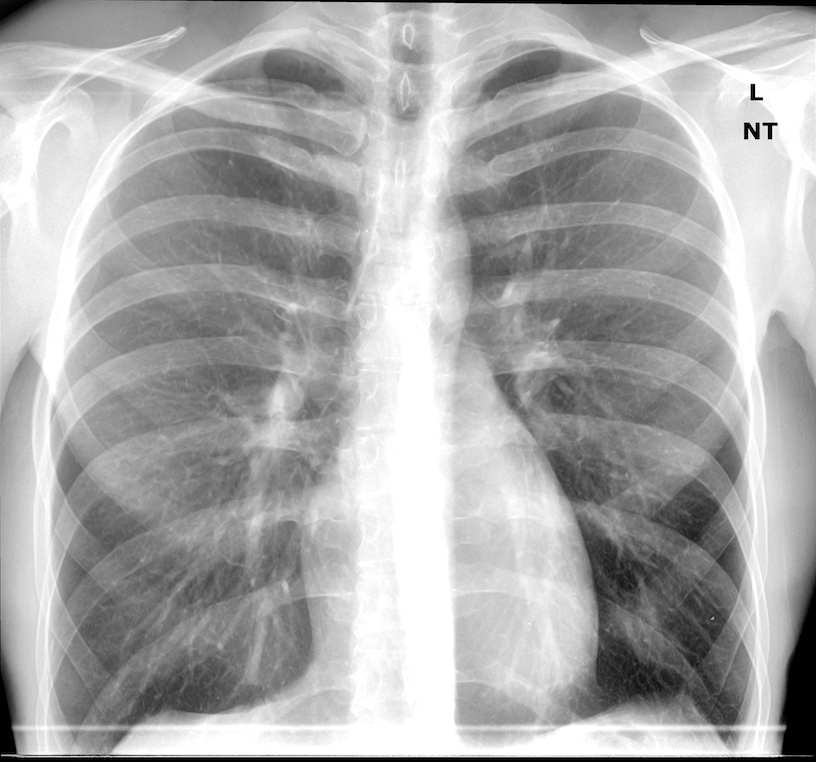
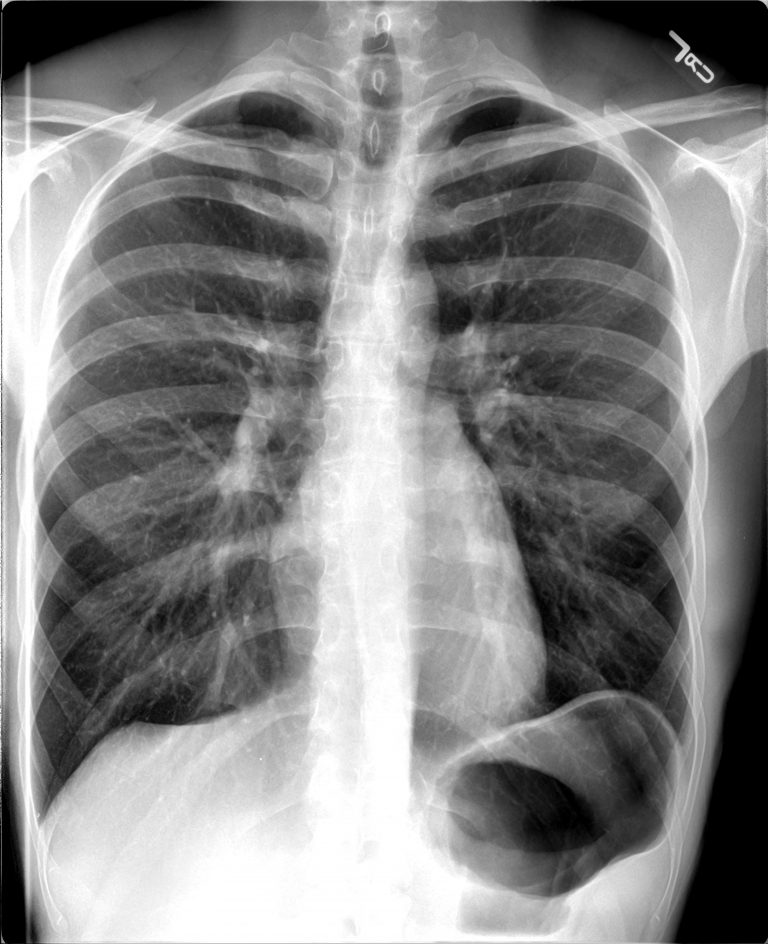
Let’s see them side-by-side for comparison. (Easier on desktop.)


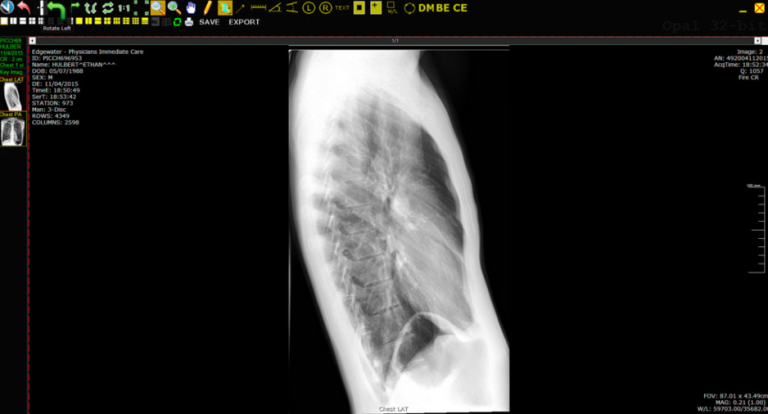
Side View Facing Right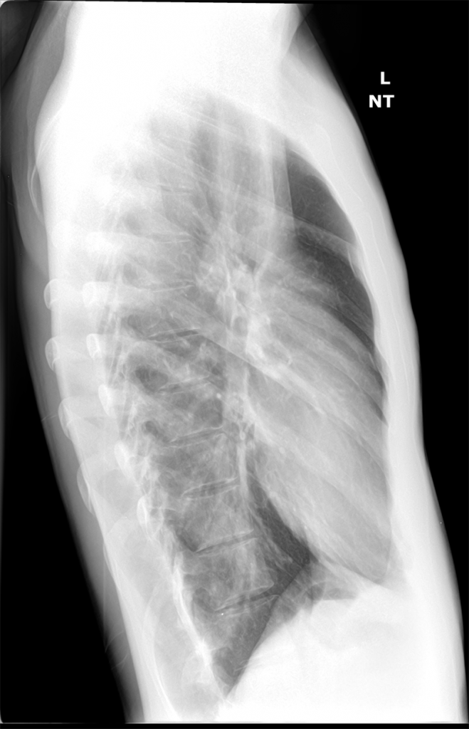

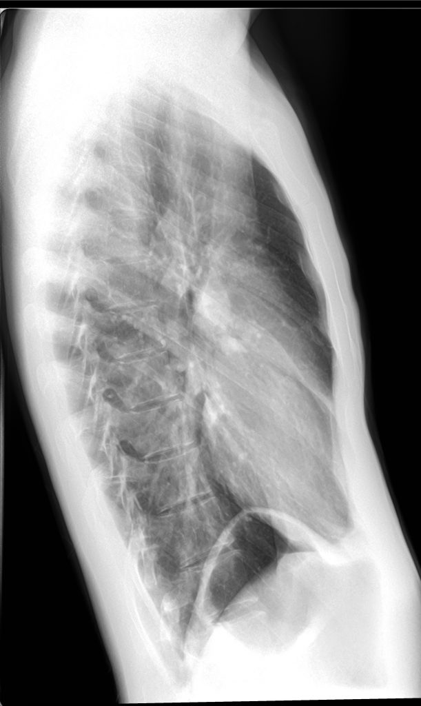
Side-by-side again:
Software View
It’s always a bonus for me when the doctors give me the full x-ray viewing software. Here’s how this hospital views x-rays digitally.
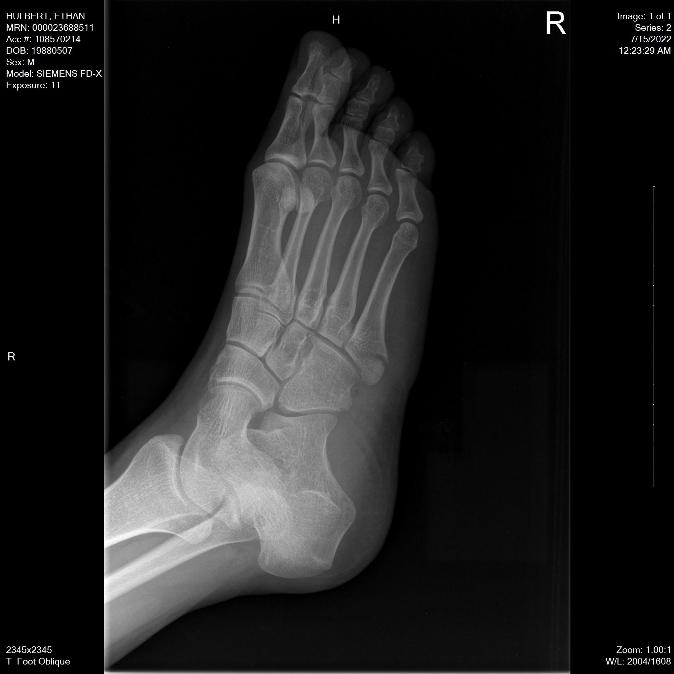
Right Foot
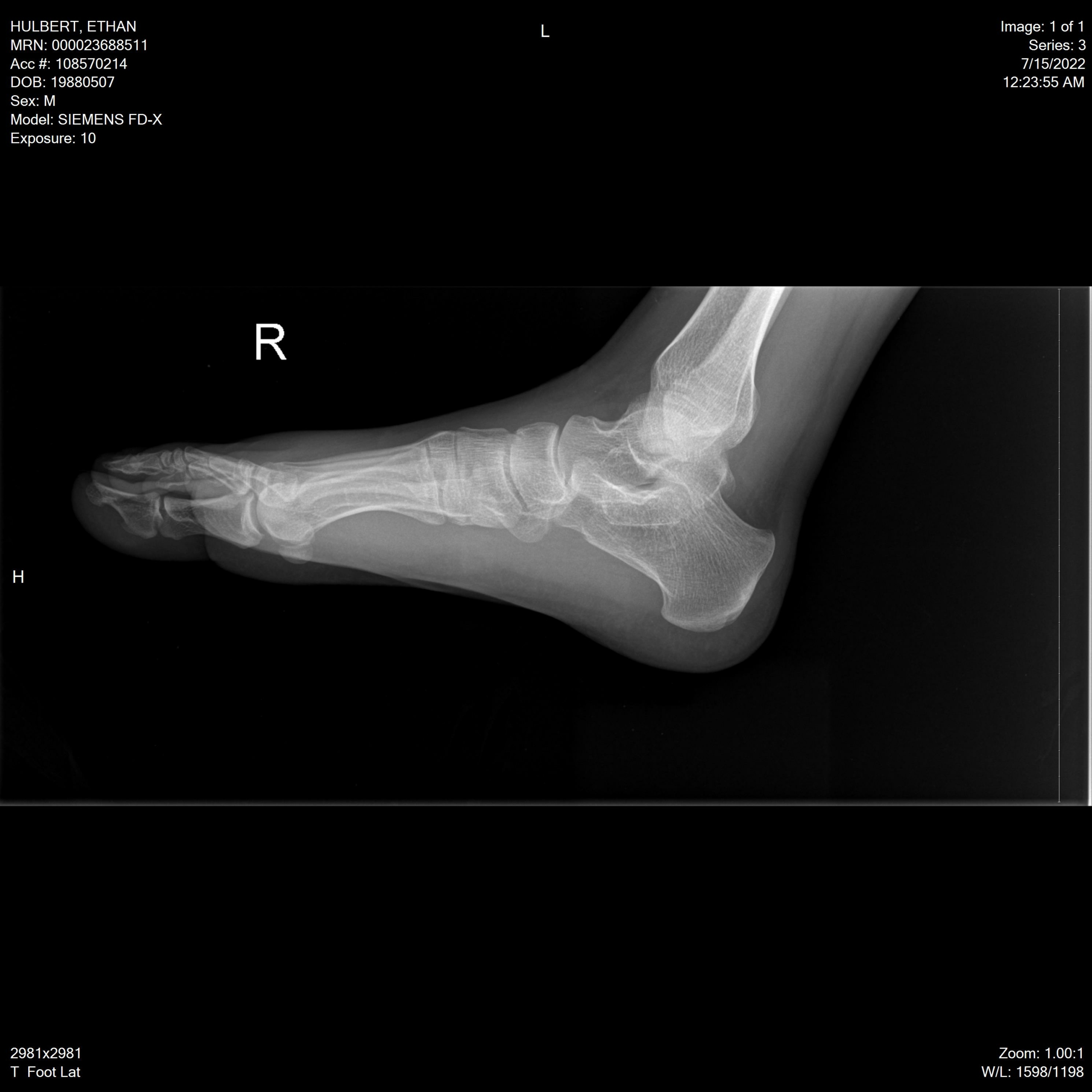
In July of 2022, I broke my right foot. I stepped over a curb wrong, twisted it, and when my weight went down on it, I’m lucky that the muscle didn’t break–instead, it was strong enough to pull one of my bones so hard it cracked it instead.
Kaiser Permanente’s service was terrible, as usual, and I was actually turned away from one urgent care facility completely before resorting to an emergency room just to get a doctor to look at it at all, although the first doc I got gave me completely wrong advice that actually hindered my recovery. Thanks, American healthcare.
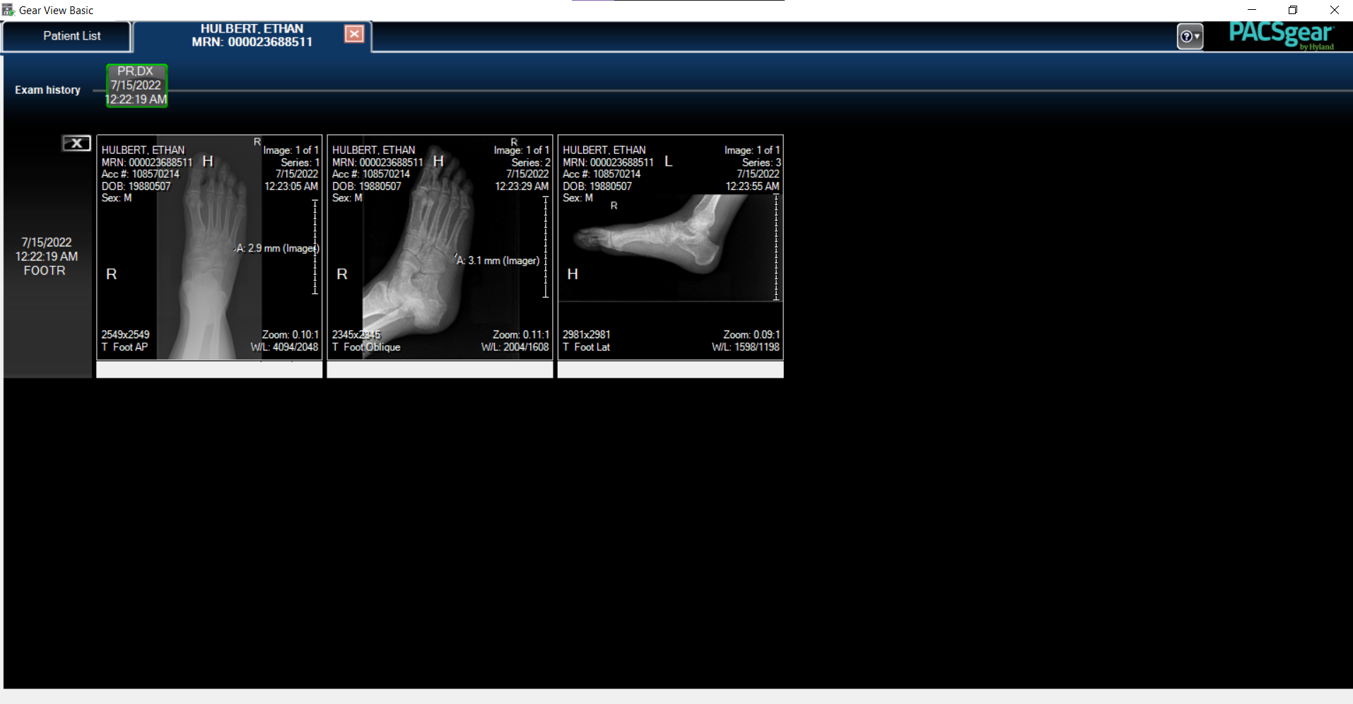
On the positive side, I got x-rays taken of my right foot from three different angles–the only useful thing Kaiser managed to do.
Their records department finally sent me the full disc of viewing software, too, and this is where things really got good. The software is called Gear View, and is part of the PACSgear medical imaging suite from Hyland.
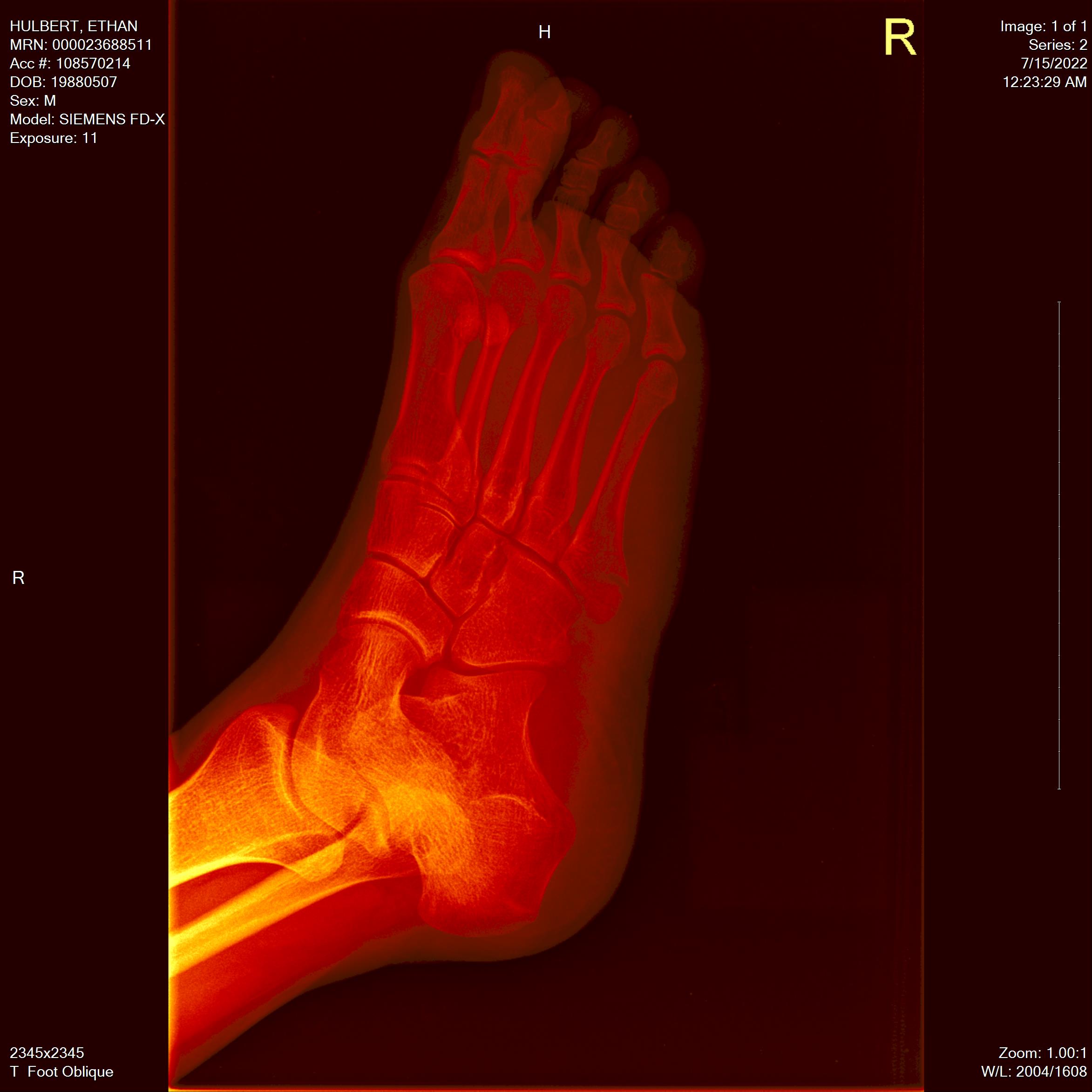
This software was fantastic. It let me see each x-ray in 7 different view modes, with different settings for brightness, contrast, color, and more. These different view modes are great for examining bones!
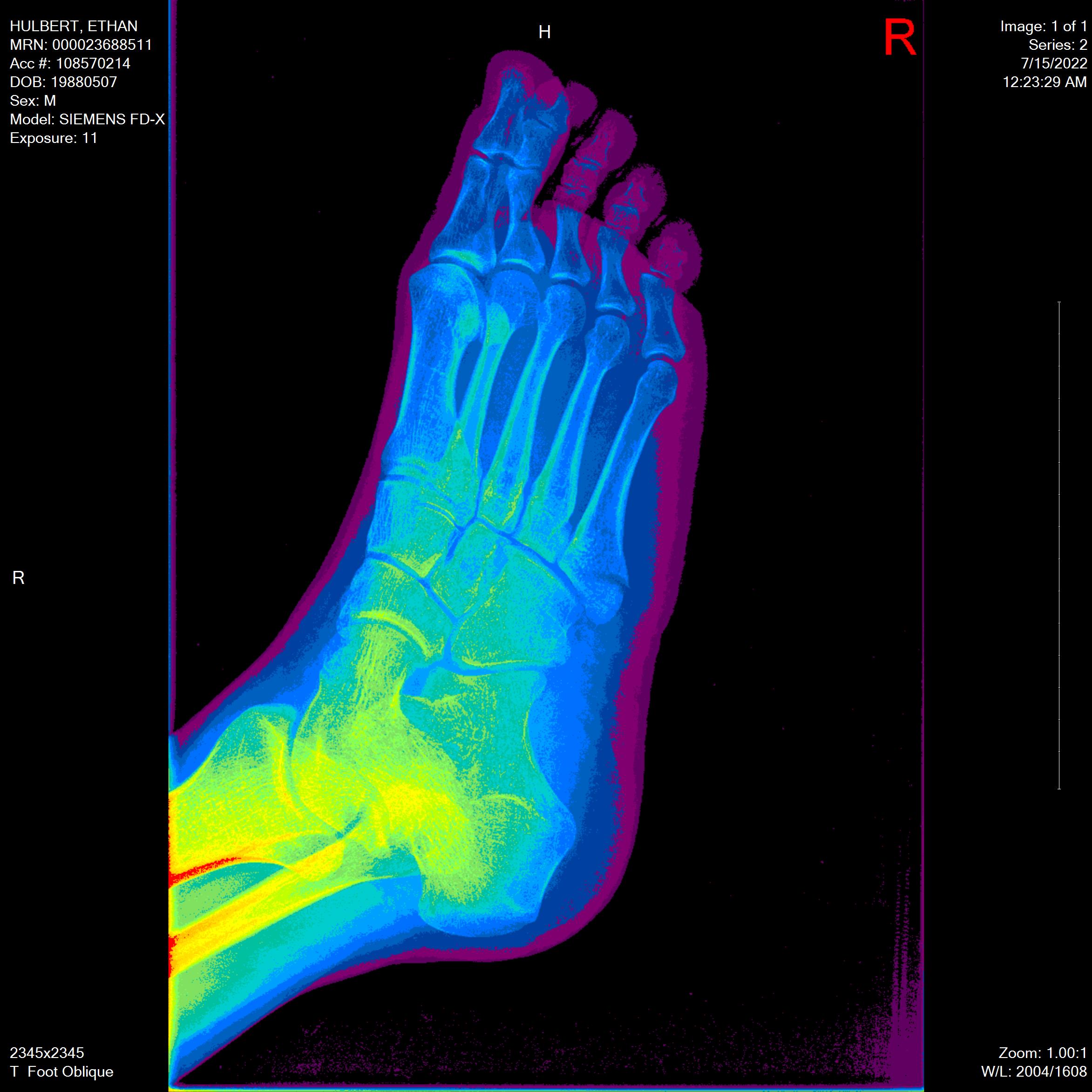
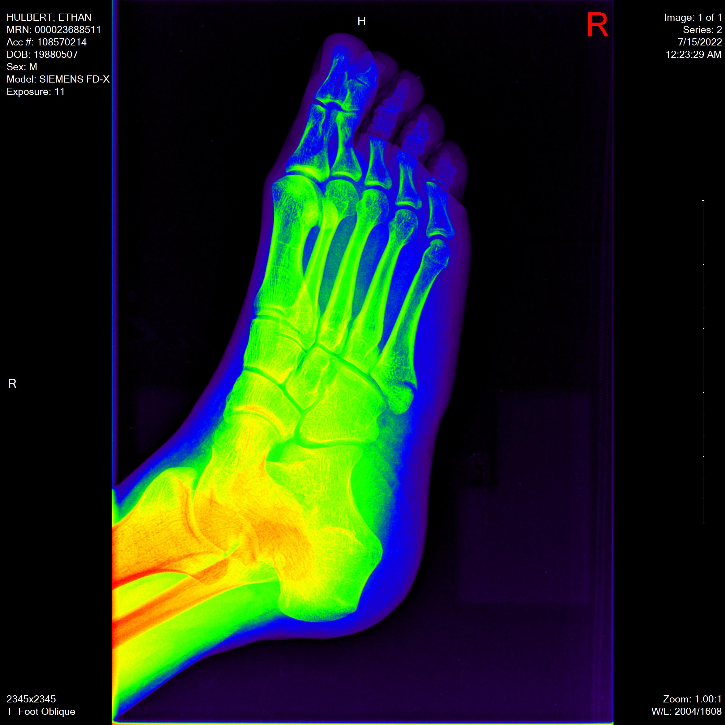
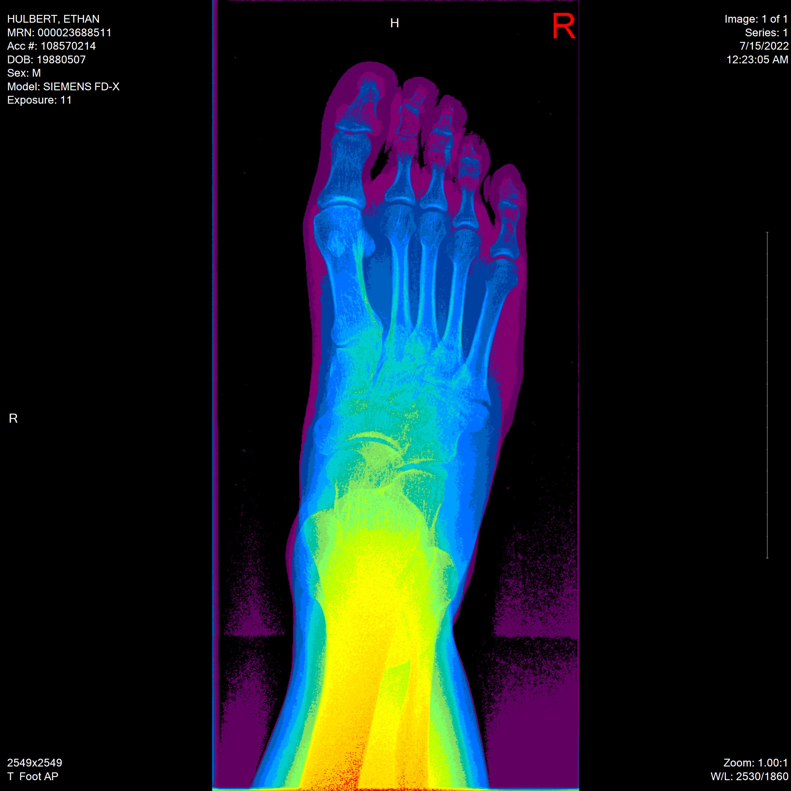
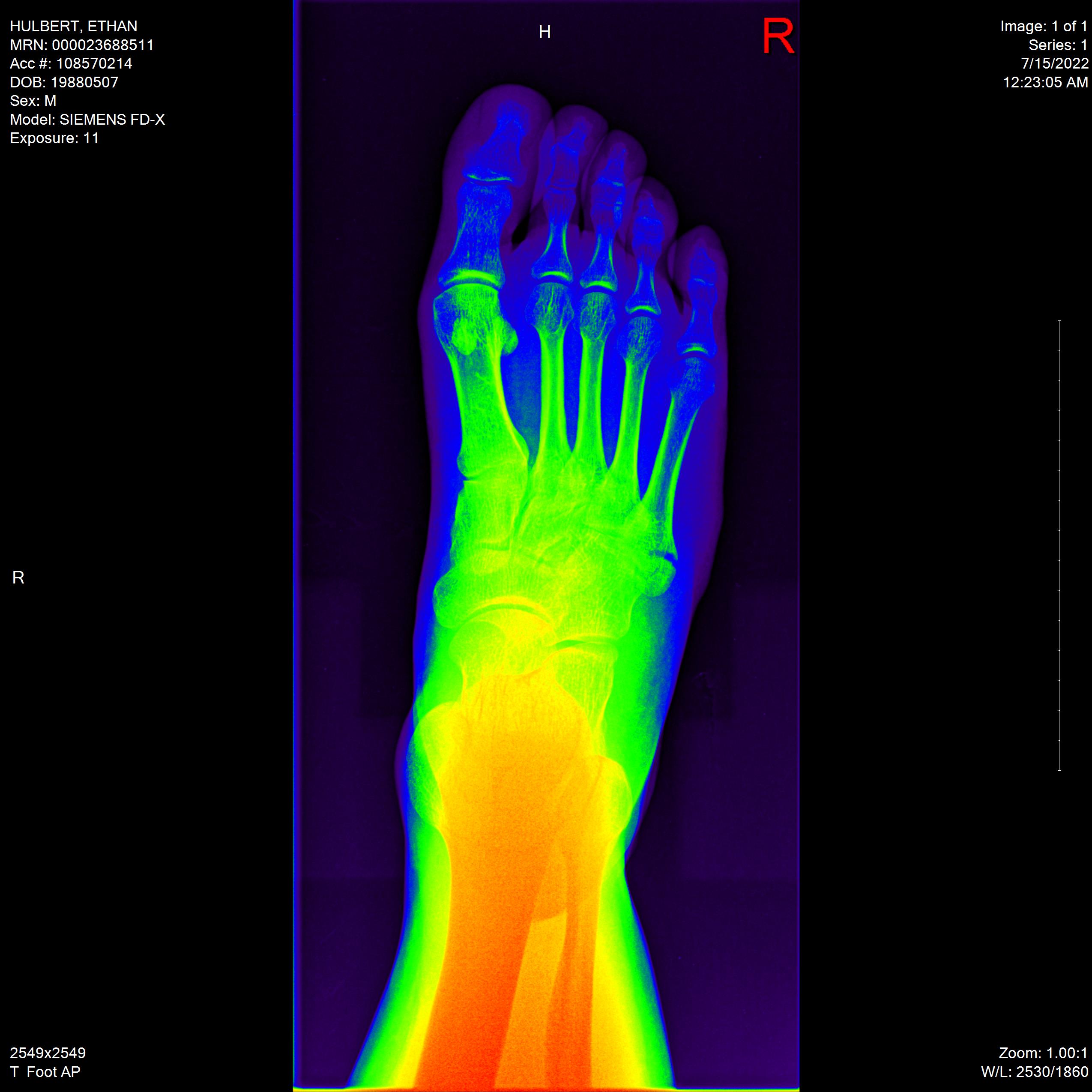
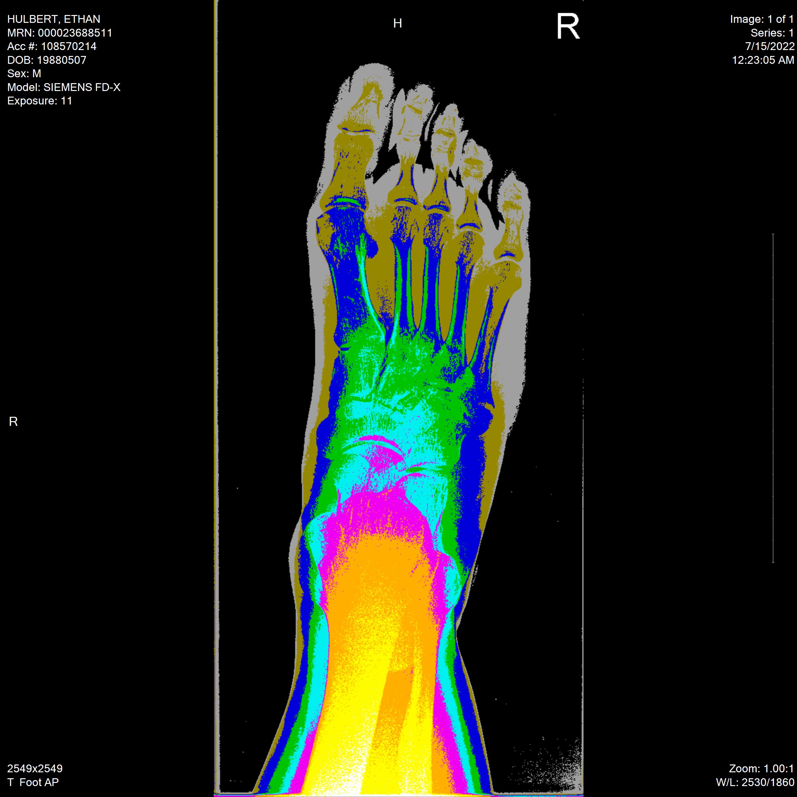
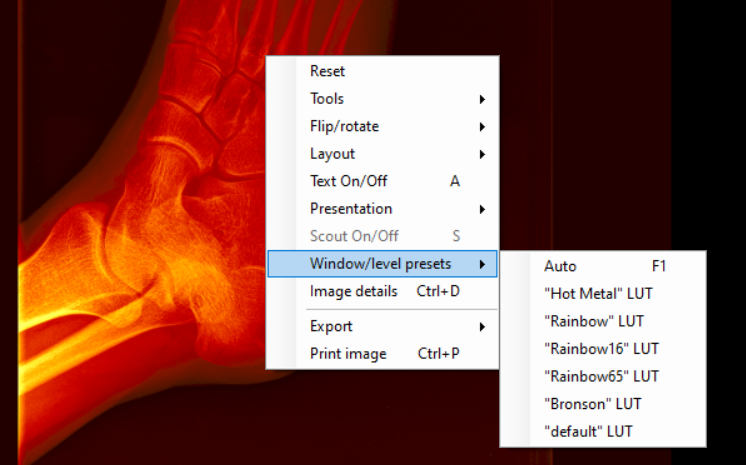
For each angle of my foot, I’ll show you the “reset” view (the default), the “auto” view (lower contrast), the “hot metal” view (molten orange–like I’m a fire elemental), three different “rainbow” modes (rainbow default, rainbow16, and rainbow65, whatever that means), and finally the “Bronson” mode. I have no idea what Bronson means.
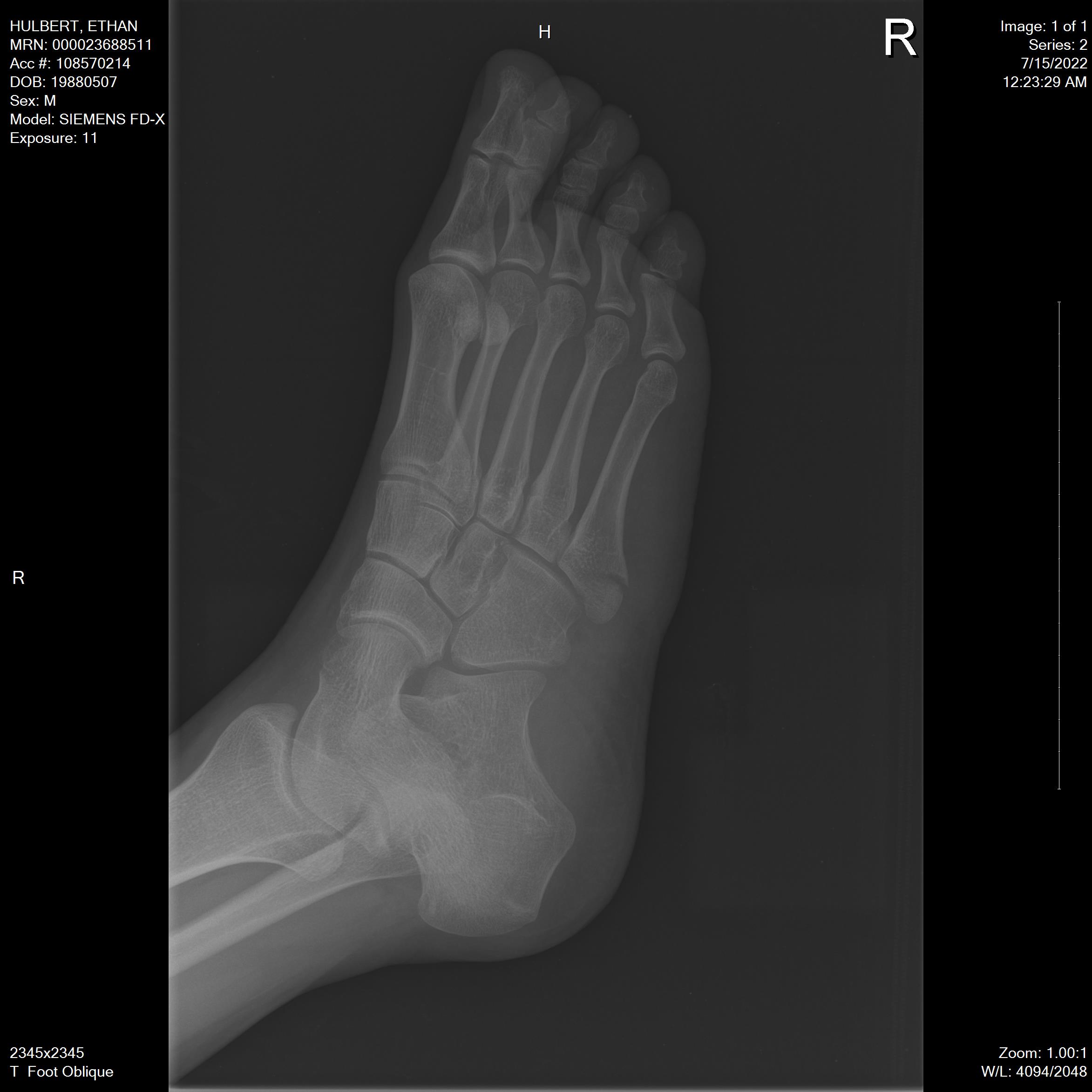
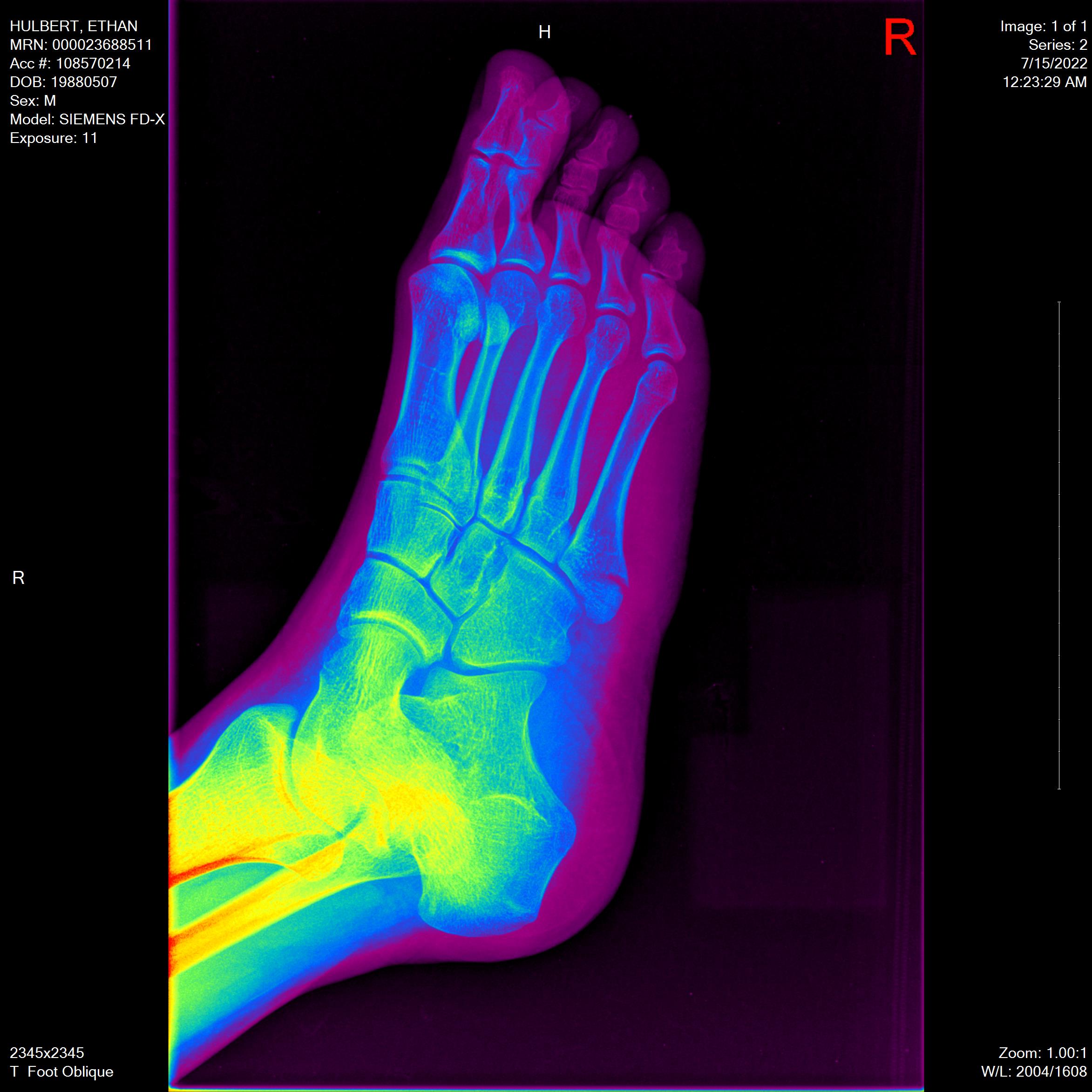
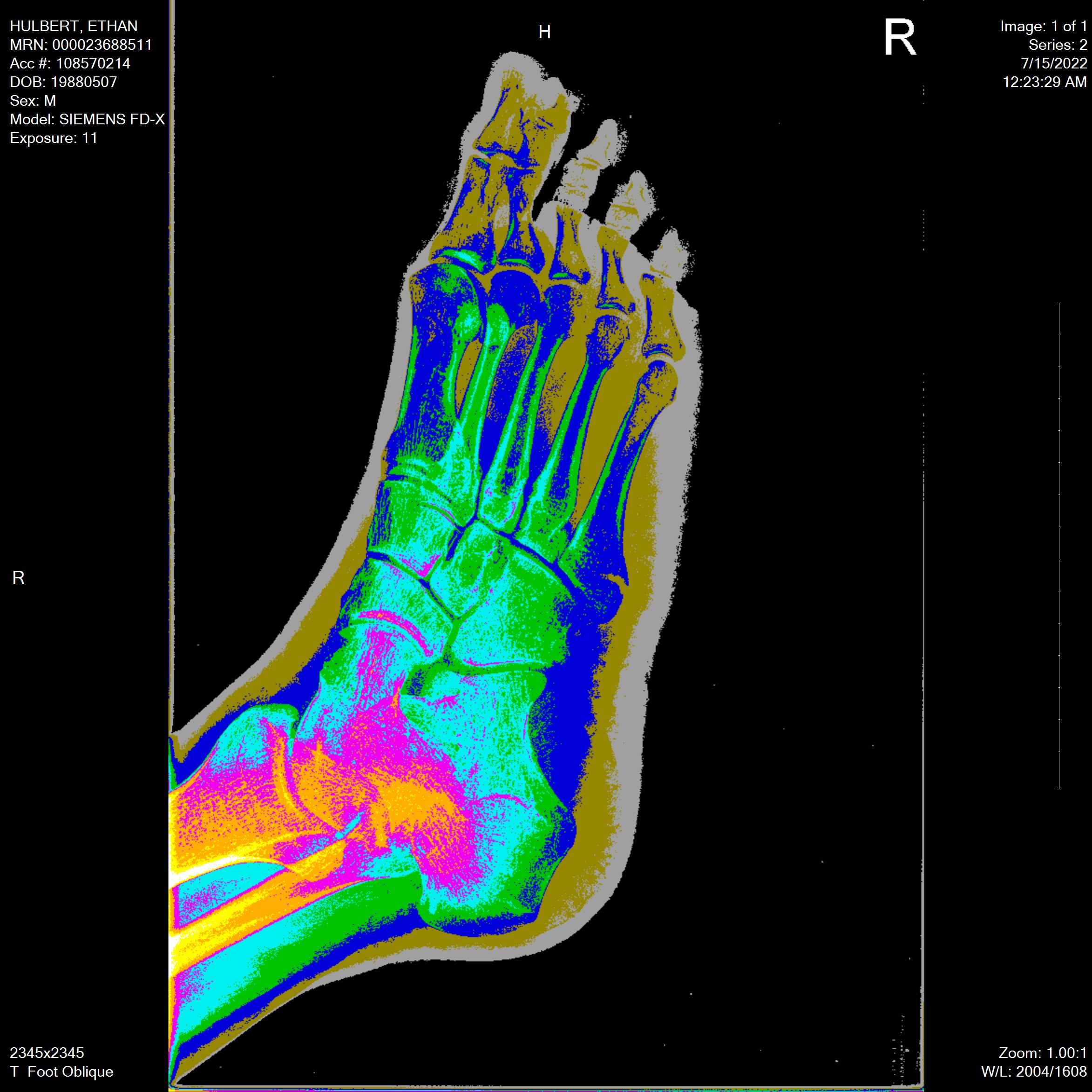
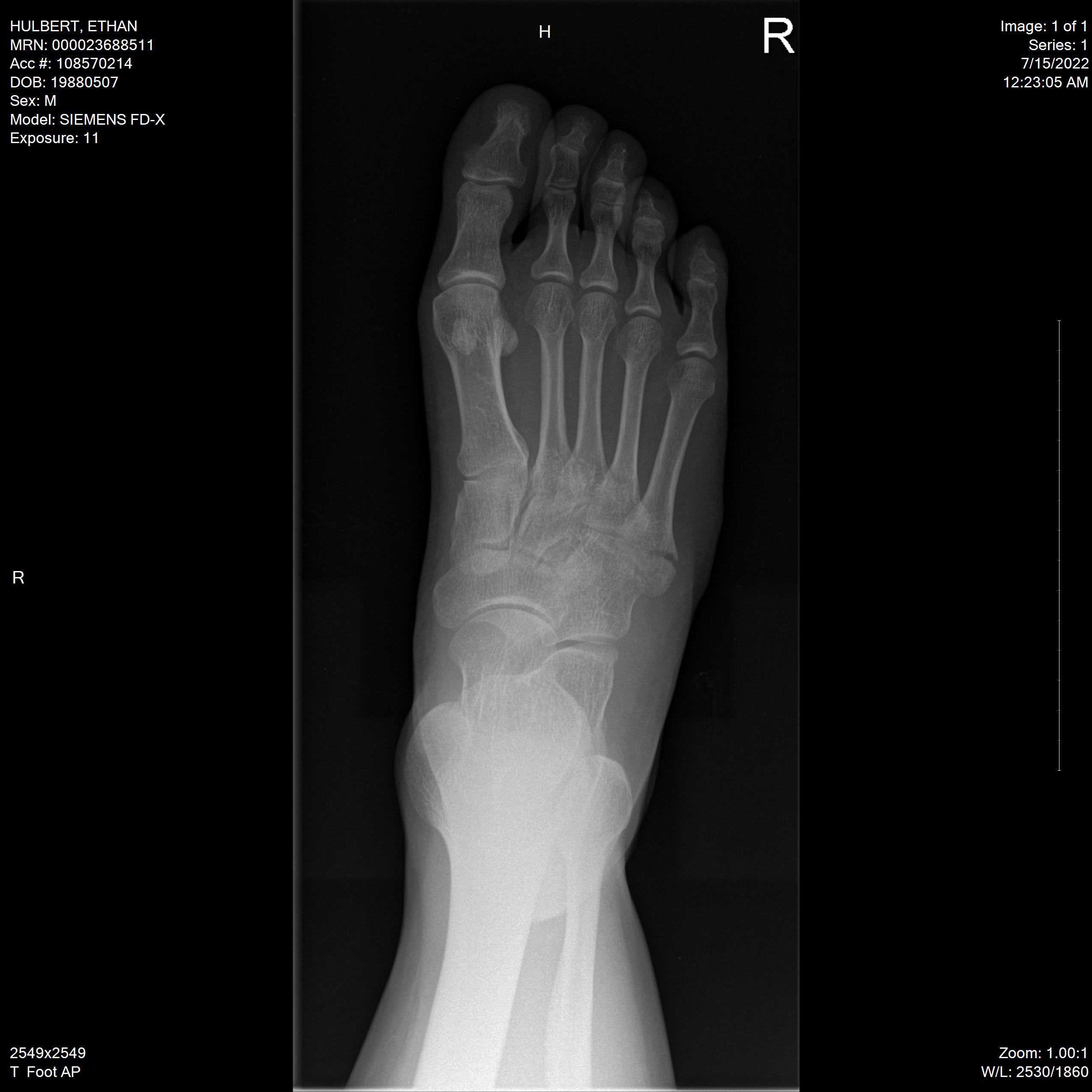
Oblique Angle
Black and White “Reset” Default View:
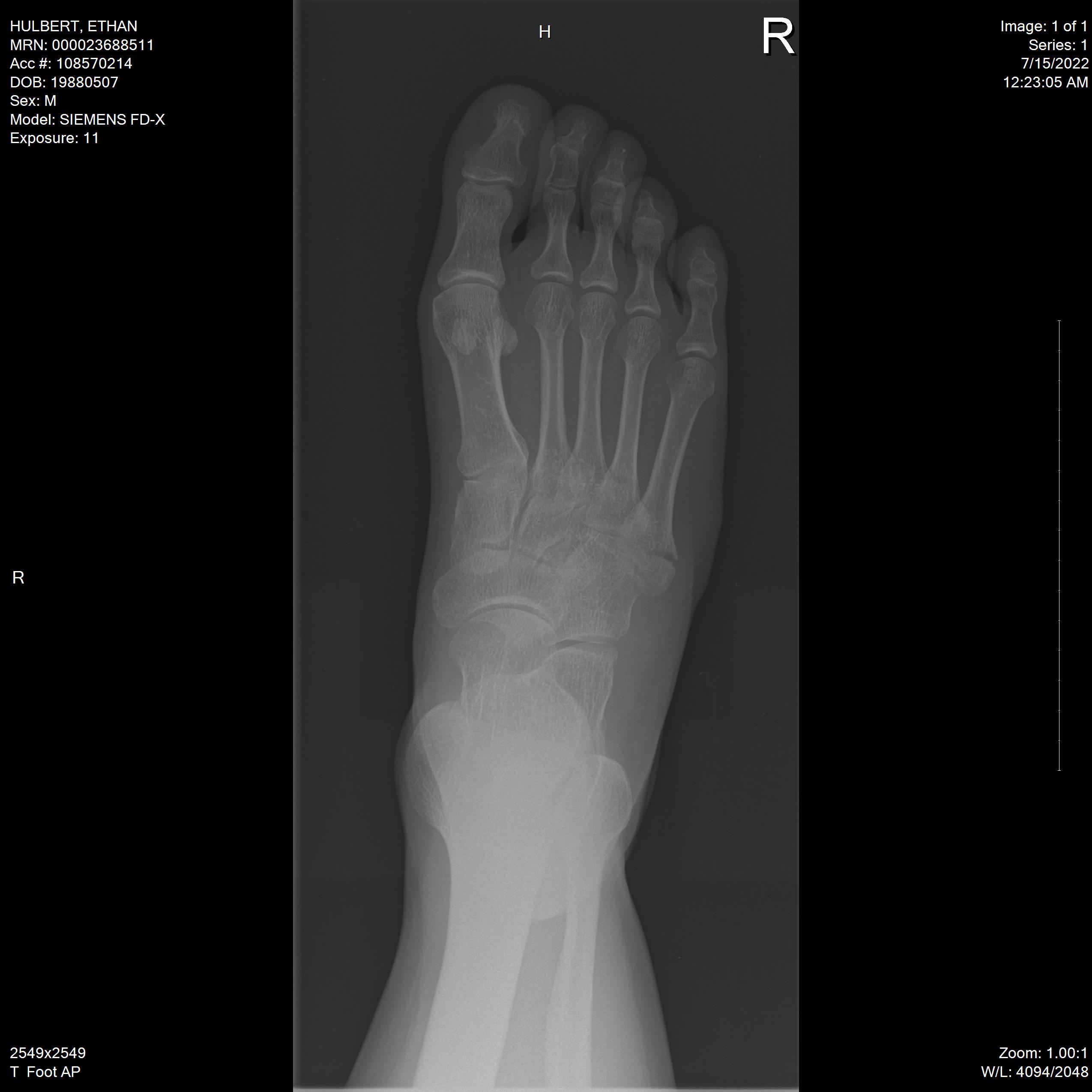
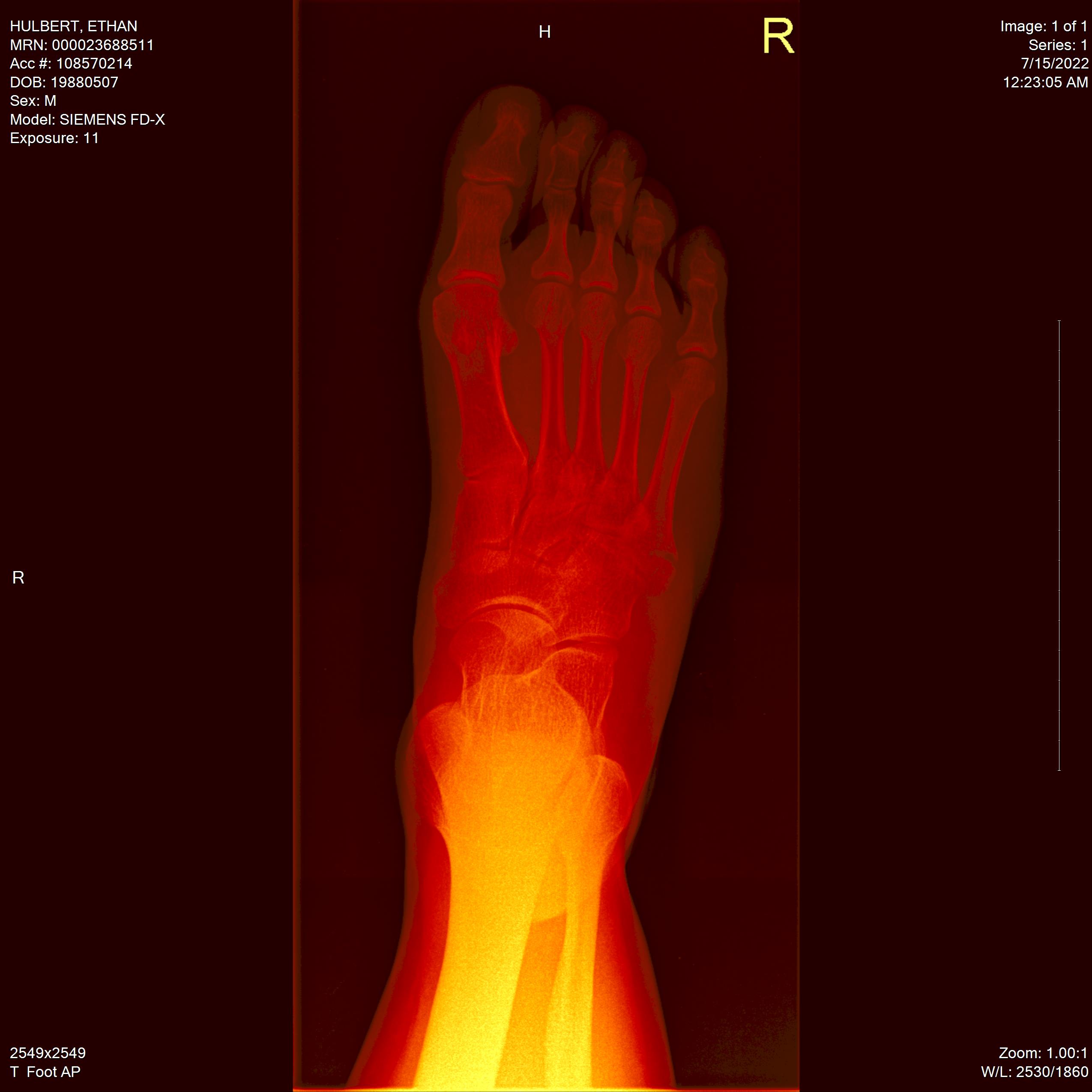
“Auto” View & “Hot Metal” View:
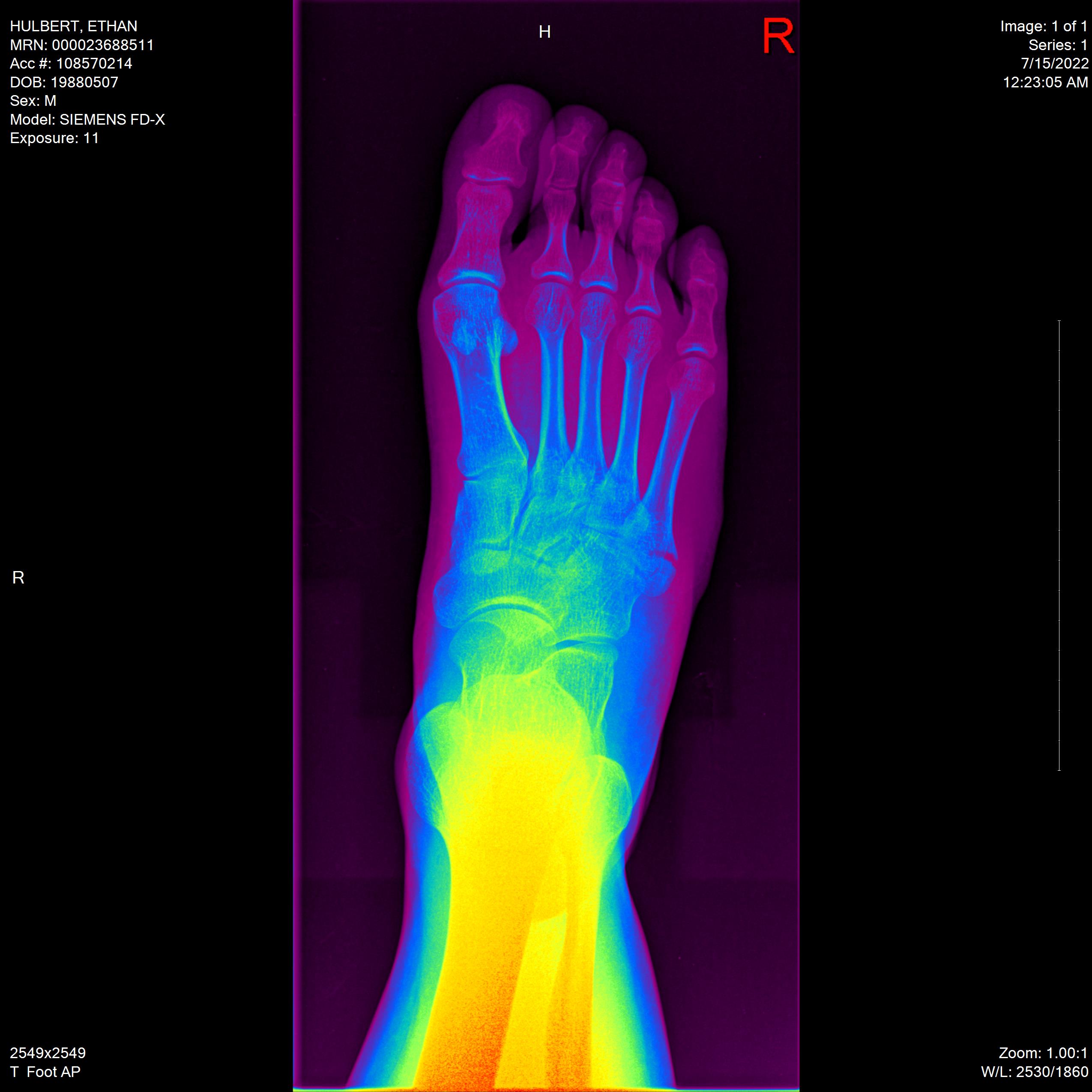
“Rainbow” View & “Rainbow16” View:
“Rainbow65” View & “Bronson” View:
From Above
“Reset” Default View:
“Auto” View & “Hot Metal” View:
“Rainbow” View & “Rainbow16” View:
“Rainbow65” View & “Bronson” View:
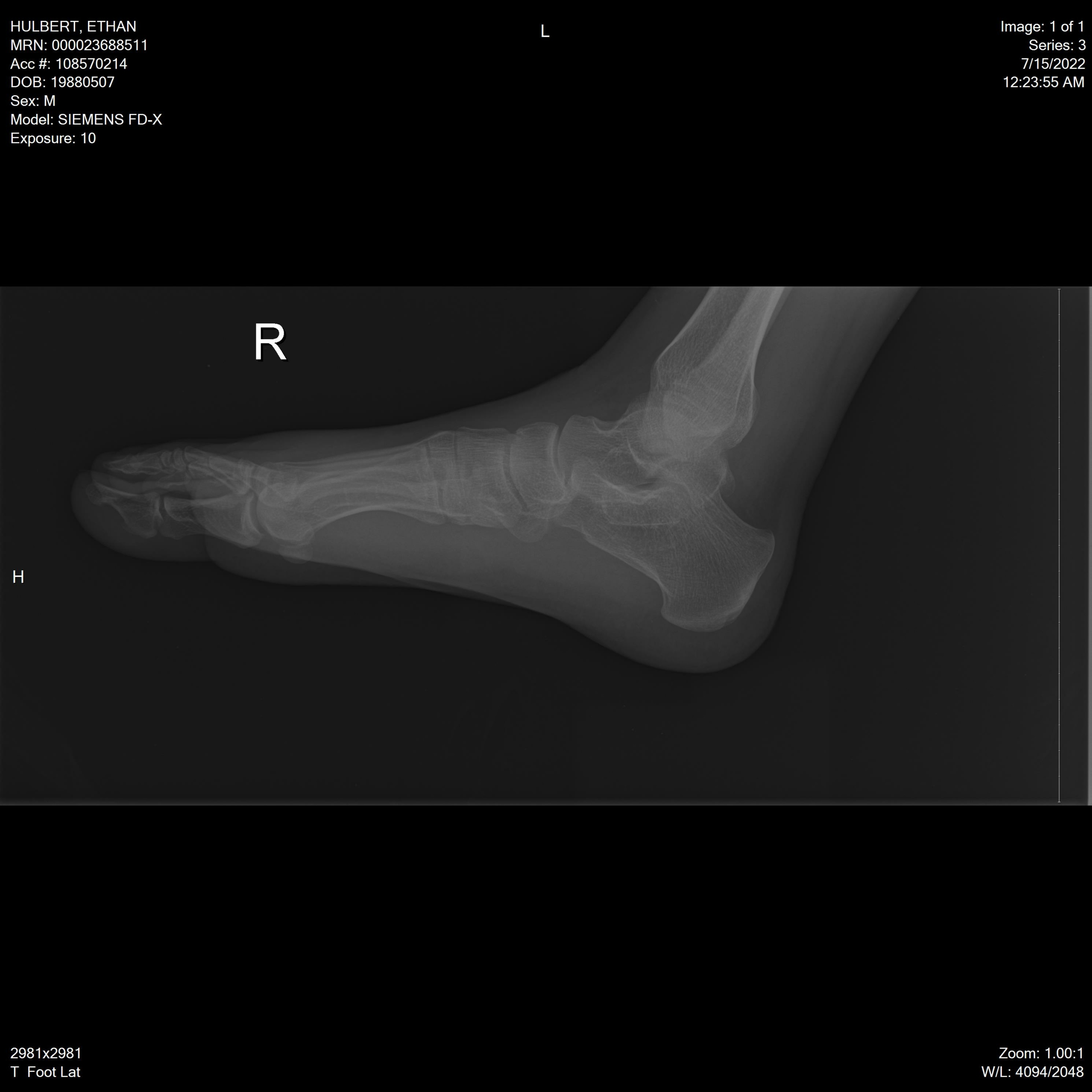
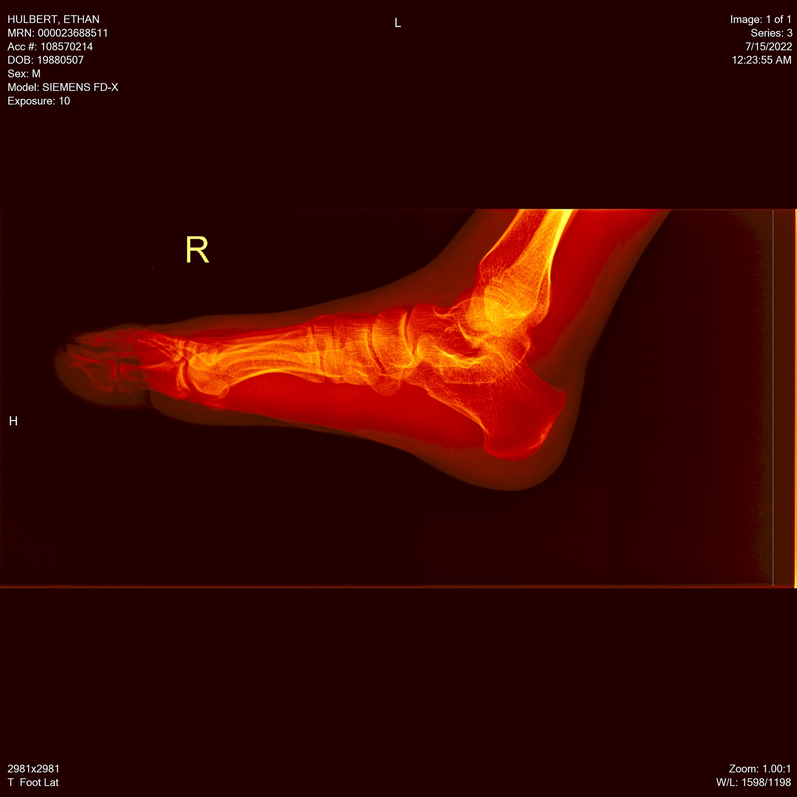
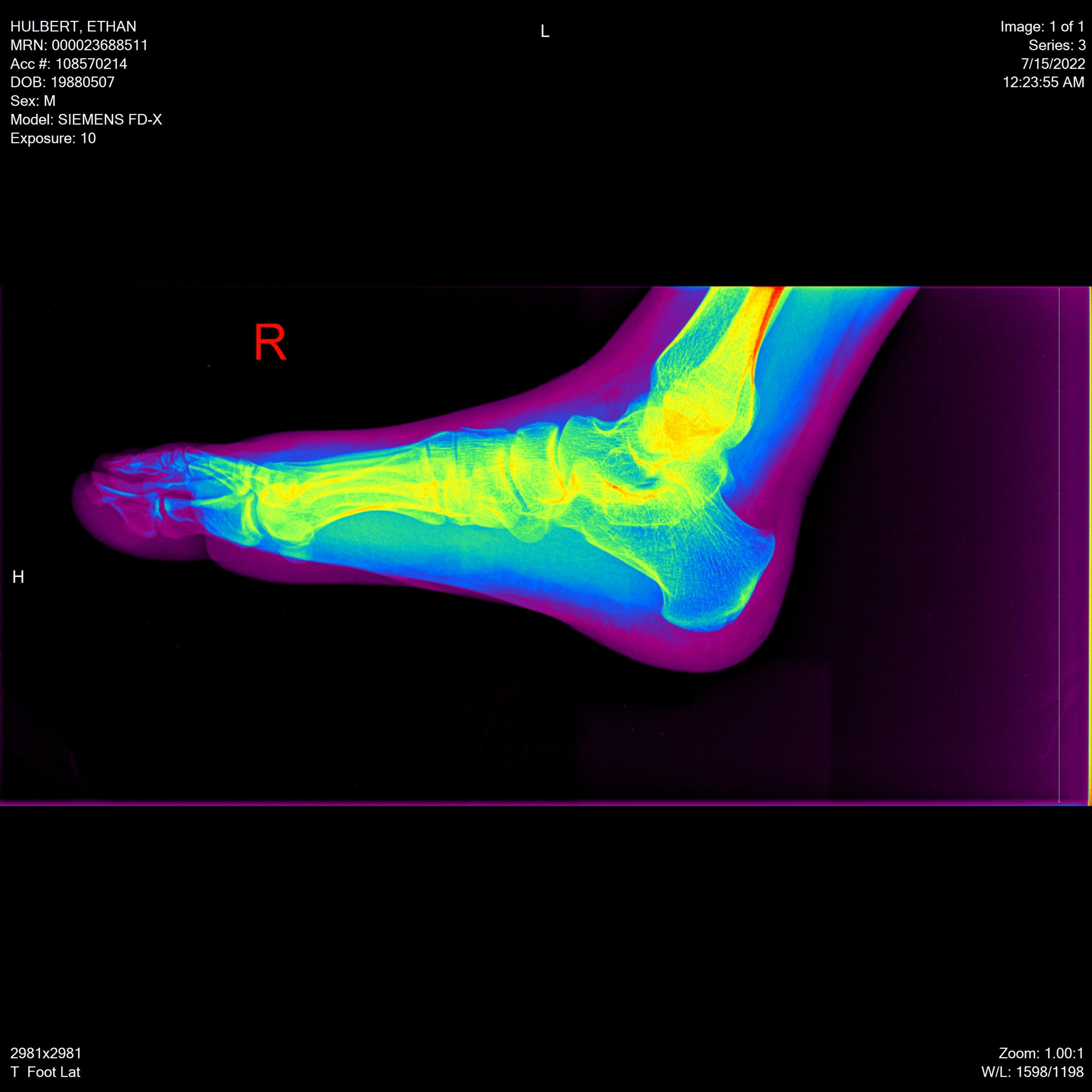
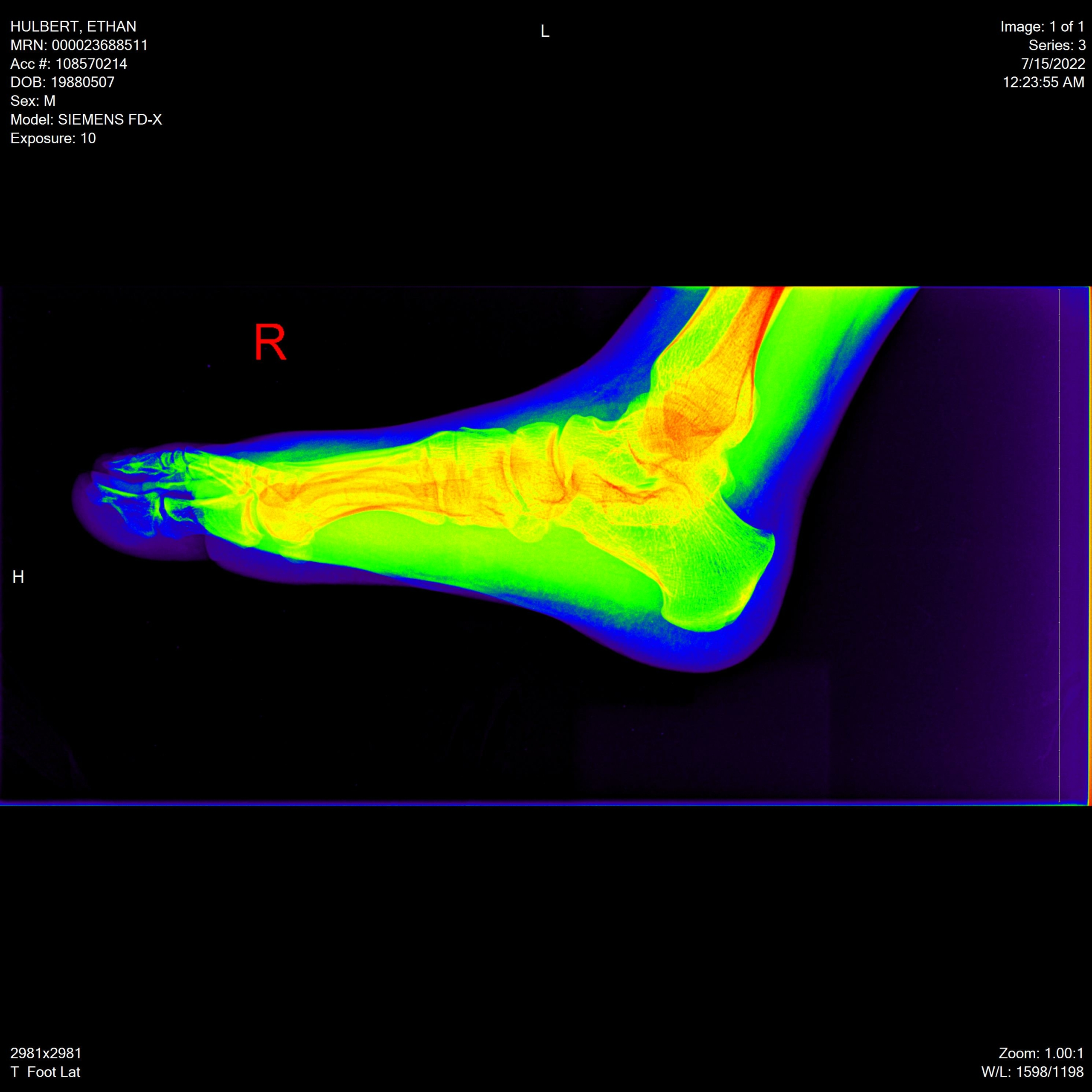
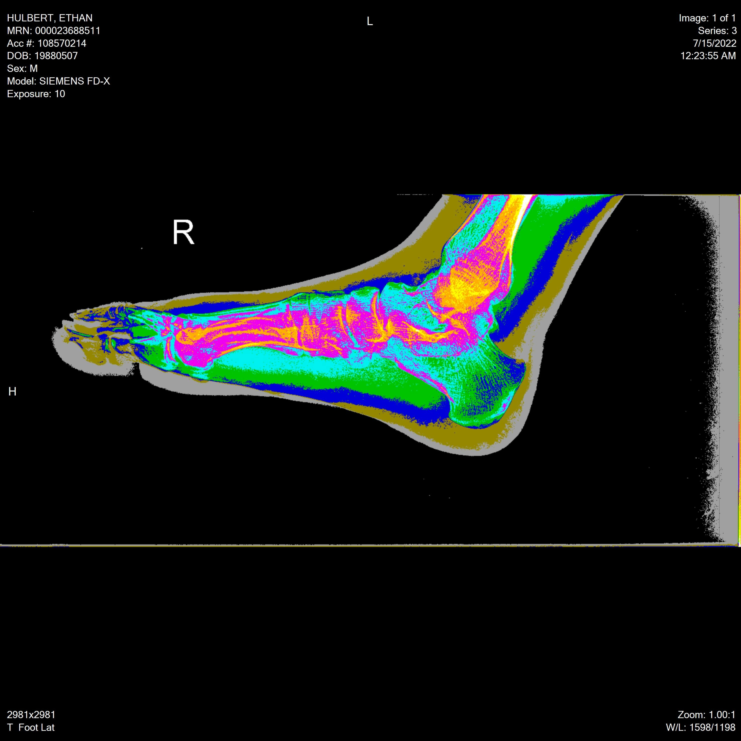
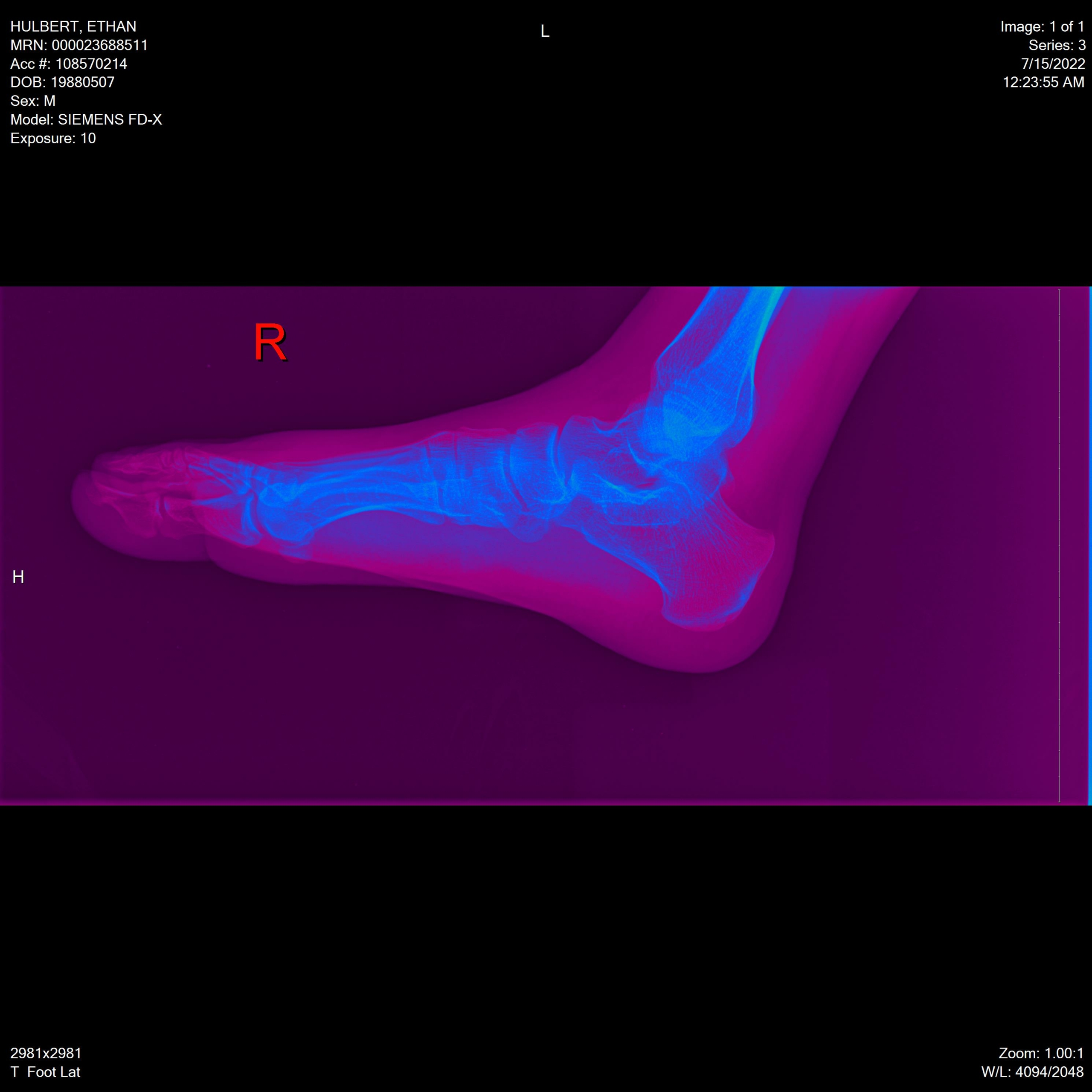
Side Lateral View
“Reset” Default View:
“Auto” View & “Hot Metal” View:
“Rainbow” View & “Rainbow16” View:
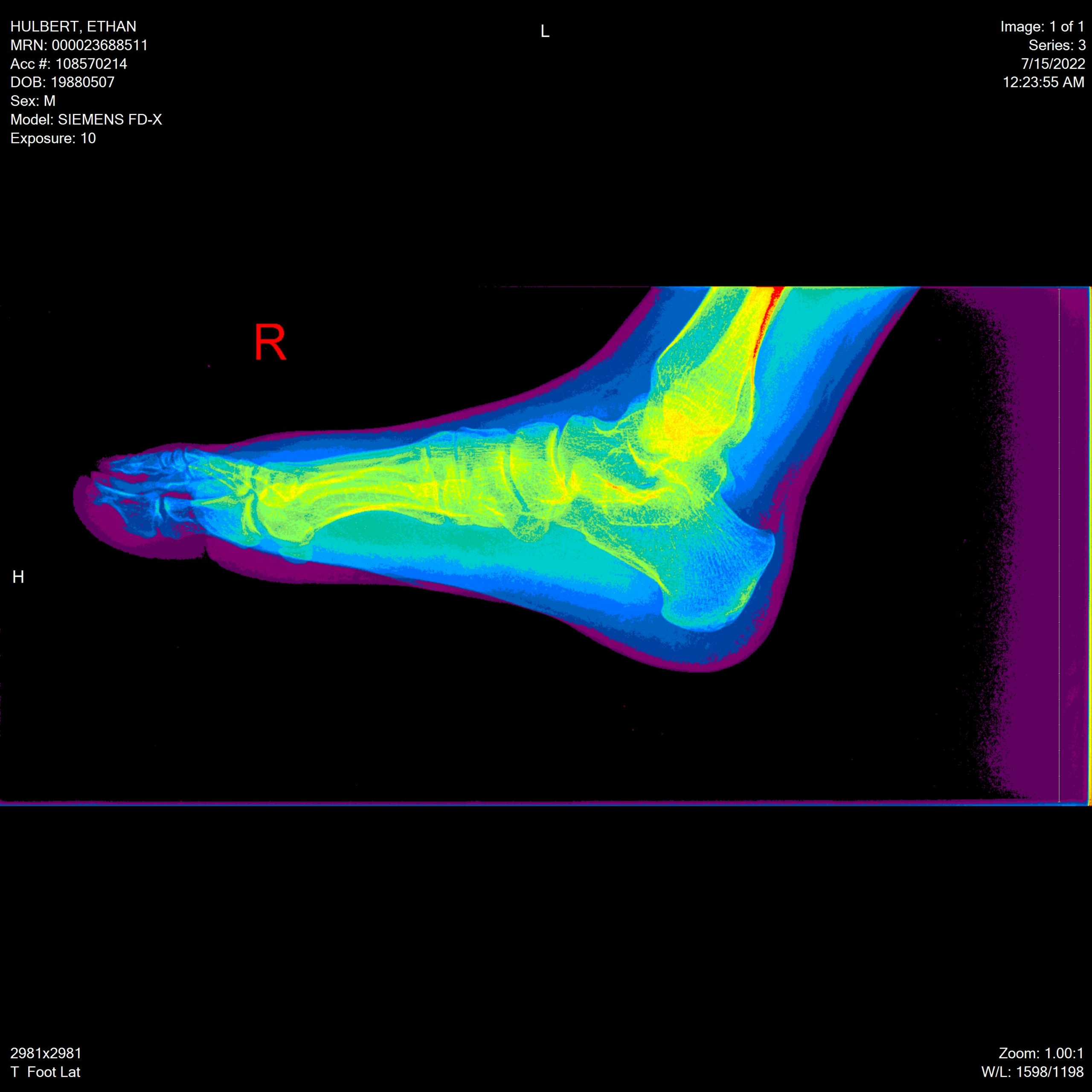
“Rainbow65” View & “Bronson” View:
I also discovered I could apply a lower contrast mode onto any of these other modes. I didn’t feel like going back and getting double screenshots for everything, but it was pretty neat. Here’s a low contrast “Auto” mode of the “Rainbow” side lateral view:
Gear View Software
The icon for the software is so freaking cute. I love this! It would have been so easy for a lazy programmer to just leave it as the default executable image, but no, someone designed this. And I appreciate it so much.
![]()
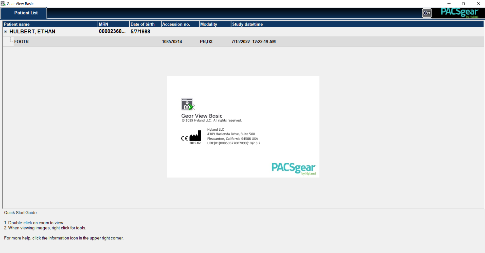
Here’s getting in to the program. You can imagine a doctor would have tons of patients with tons of x-rays all down this screen.
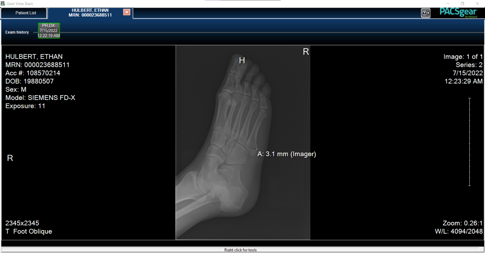
Then we can select which x-ray angle we want to drill down into.
This is the full view of a specific x-ray in a specific view. It has a little more information around it. Also, note that point A on the image is where my bone breakage occurred.
Then we can do all kinds of things to help us view the image. Here’s the menu that specifically shows me all those different view modes from above, and how I know what they’re called.
My favorite other tool lets us draw a line between two points across the x-ray, and it gives us a precise measurement! I can imagine that this would be extremely helpful for doctors and nurses. I wonder how the software matches the reference scale to the range of depth of the image that’s in focus?
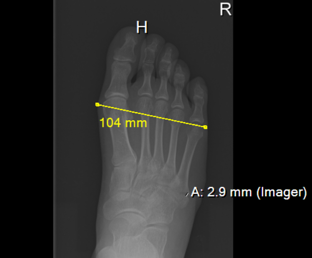
You can again see where my bone was pulled apart and separated by about 3mm, too. That’s the distance of the gap. Very cool (but ouch).
I really appreciate Hyland and the Gear View software, and I hope that when I break all the rest of my bones in the future I get the same level of convenience for viewing them.
Teeth
Many people think that dental x-rays aren’t as exciting as those of other body parts. I don’t think that could be further from the tooth.
Who knows? If I ever go missing and a disfigured body is found, these could help identify me.
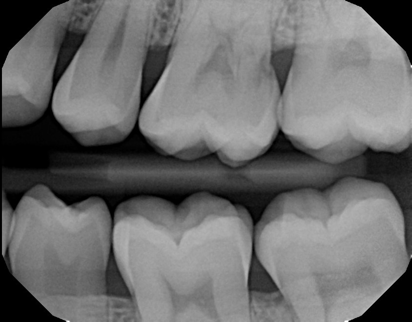
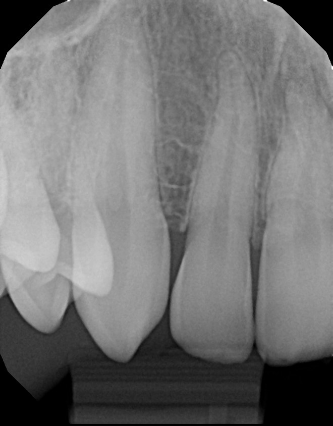
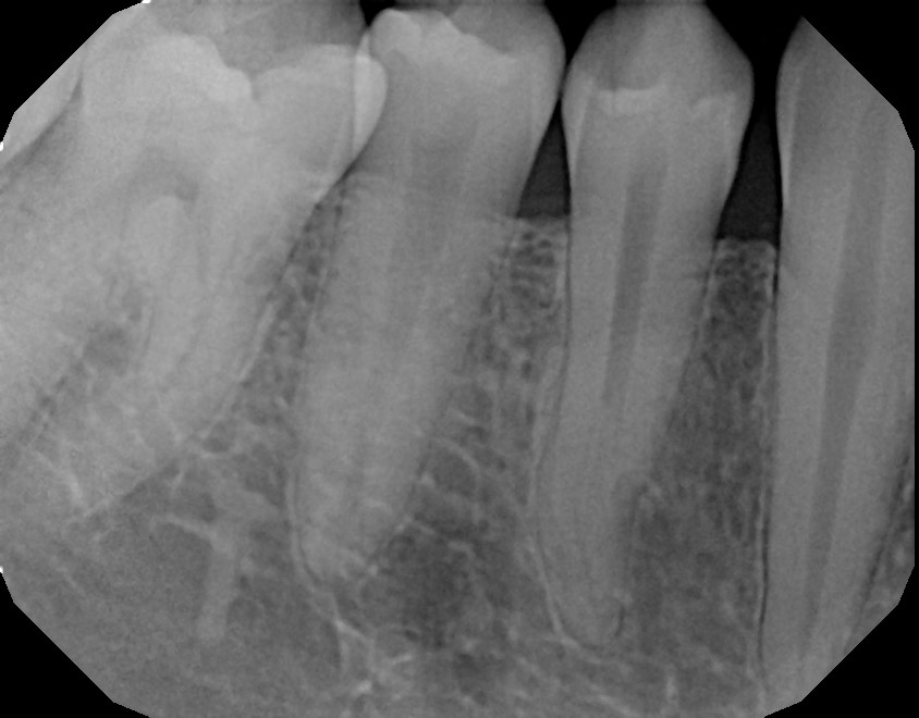
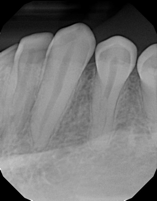
2024 January
From Victoria Olshansky D.D.S. in West Hollywood.
I went in for a toothache and found out it was due to tooth decay. Whoops. I needed a good amount of numbing and drilling after this.
I was only given the exported image as a jpeg file, but it came with this other data: “SOTA Image Export, Layout: 1PA.”
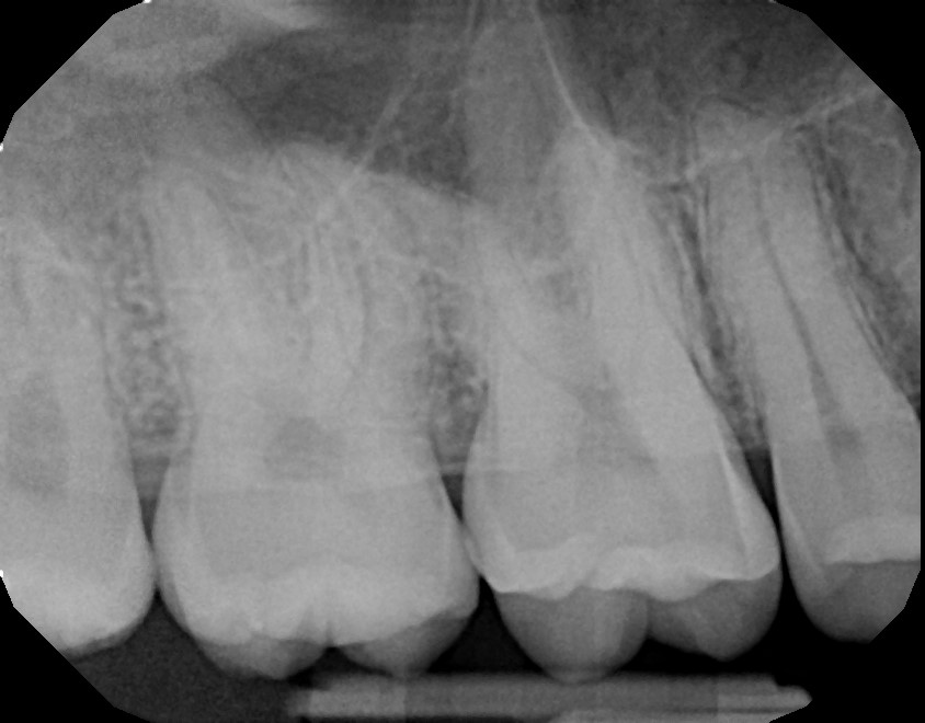
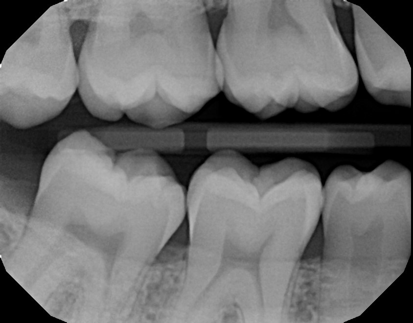
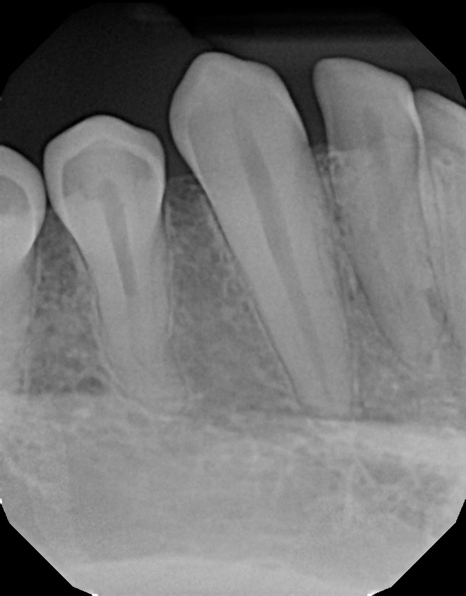
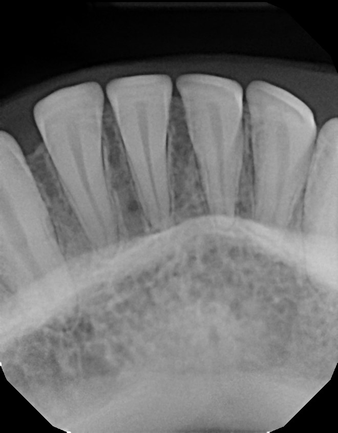
A week and a half later, I went back for the longer portion of the procedure, and got this:
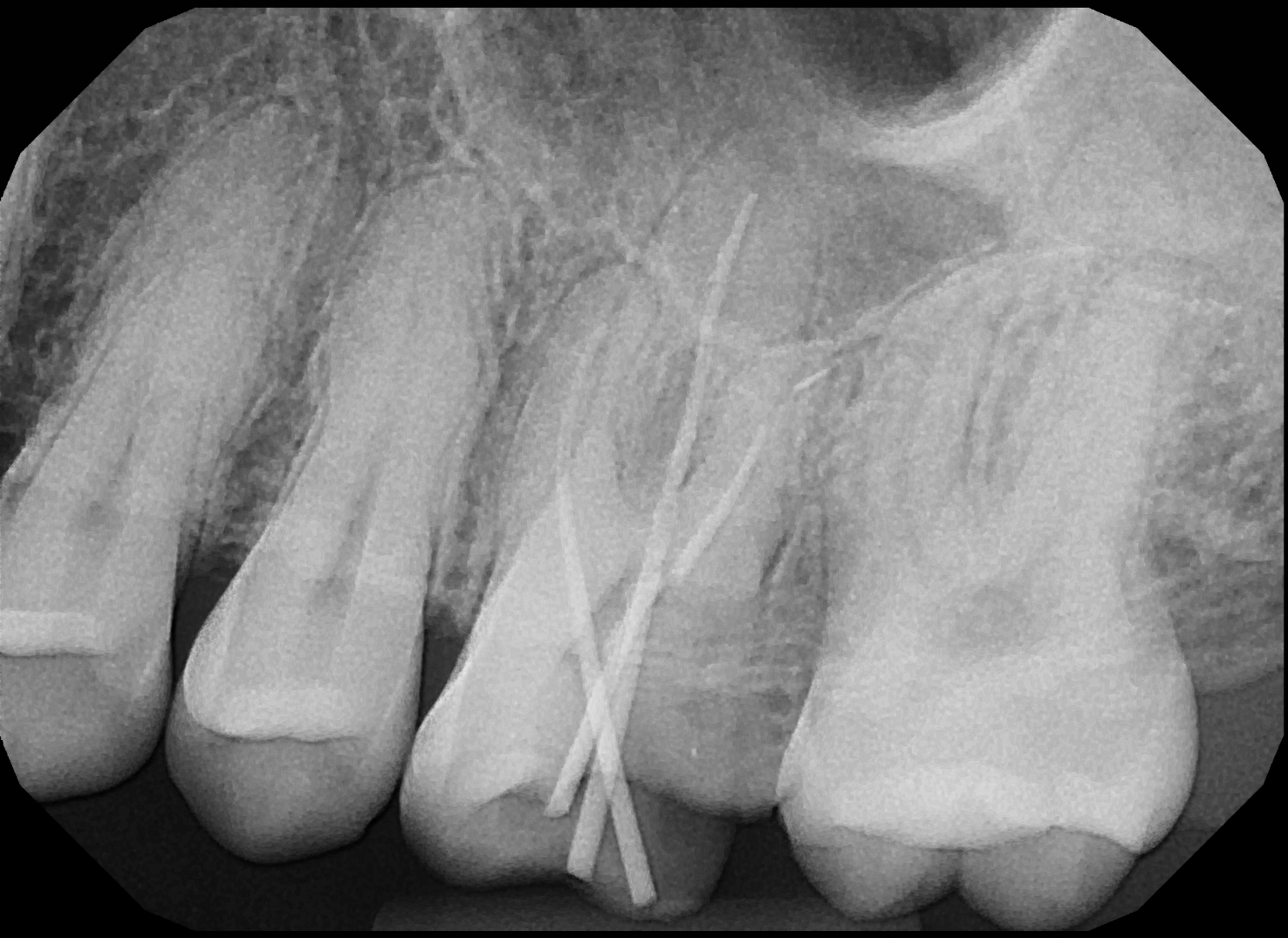
Yikes!
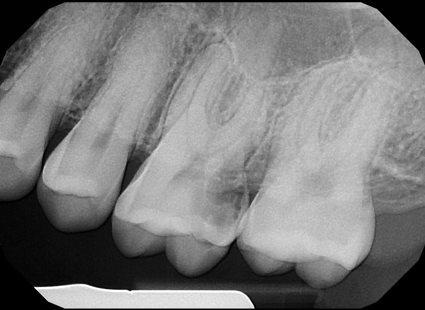
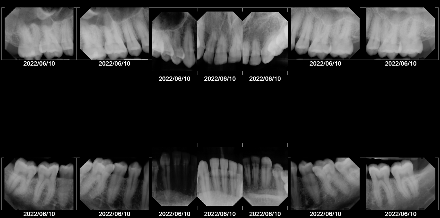
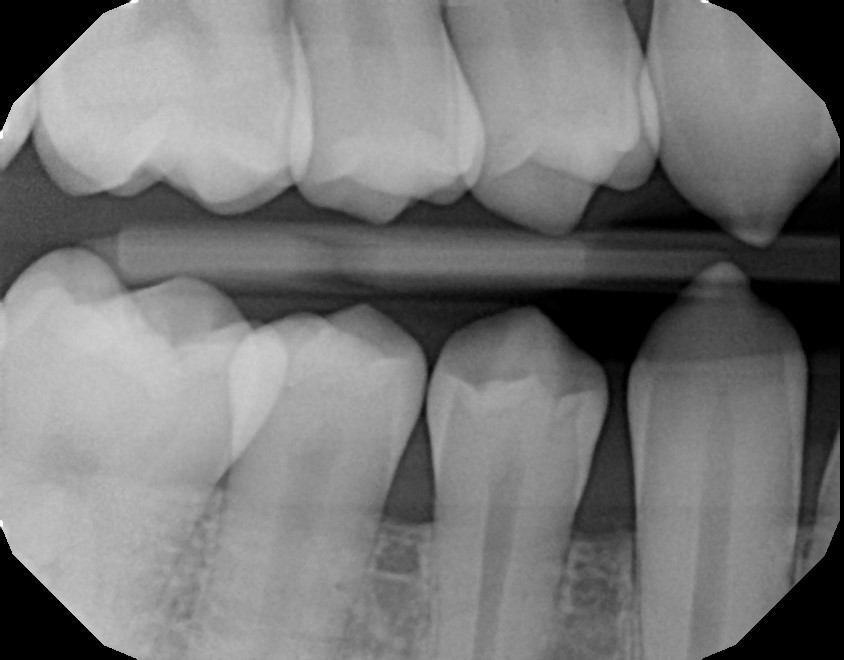
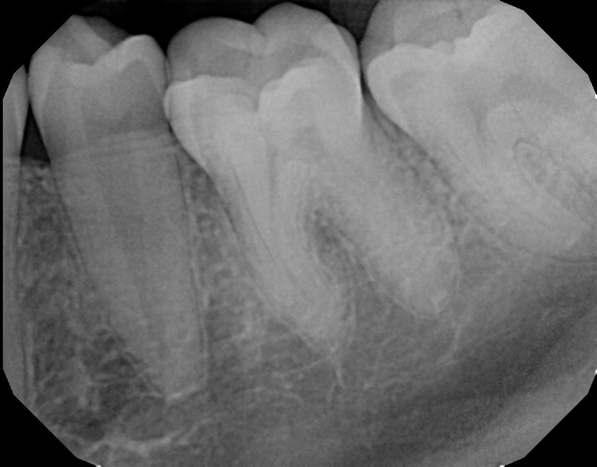
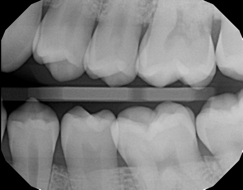
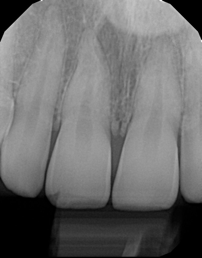
2022 June
From Victoria Olshansky D.D.S. in West Hollywood.
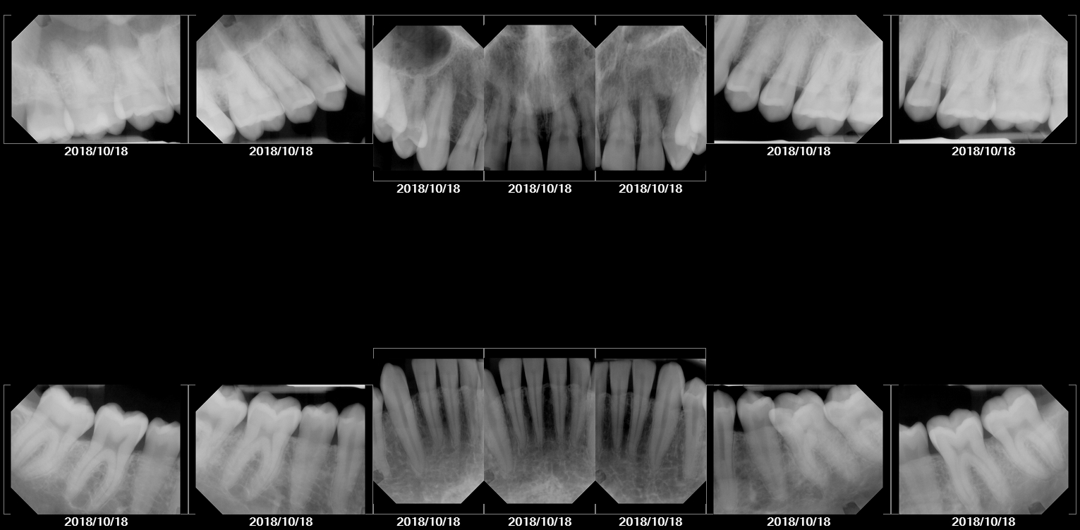
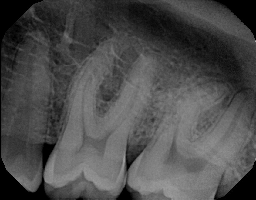
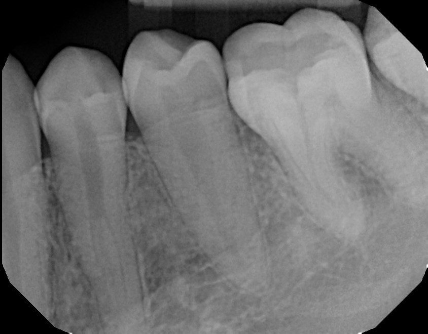
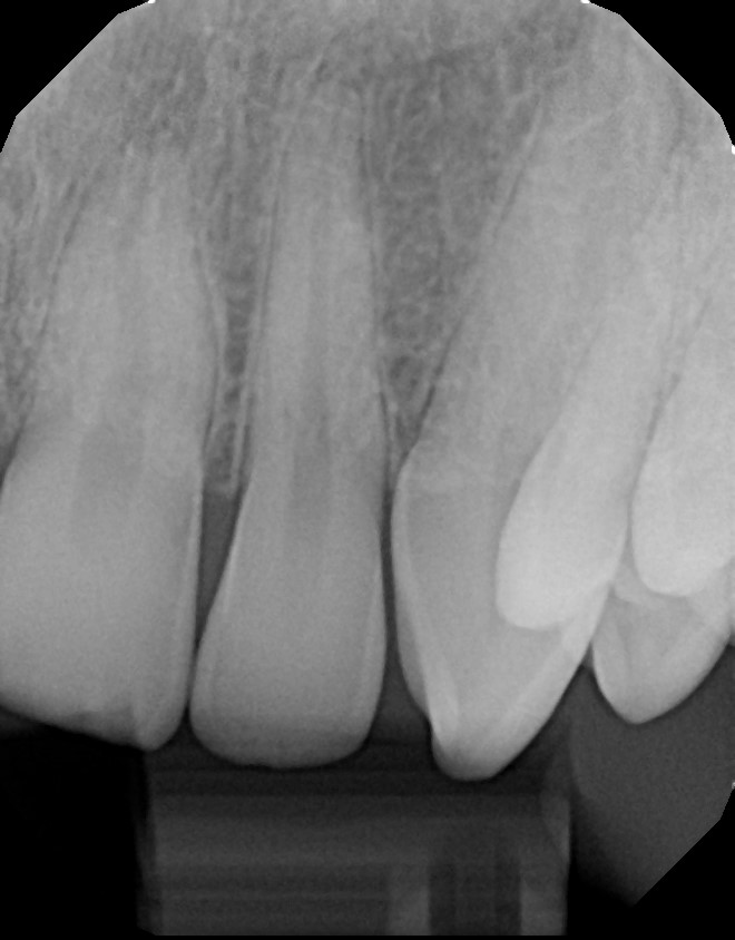
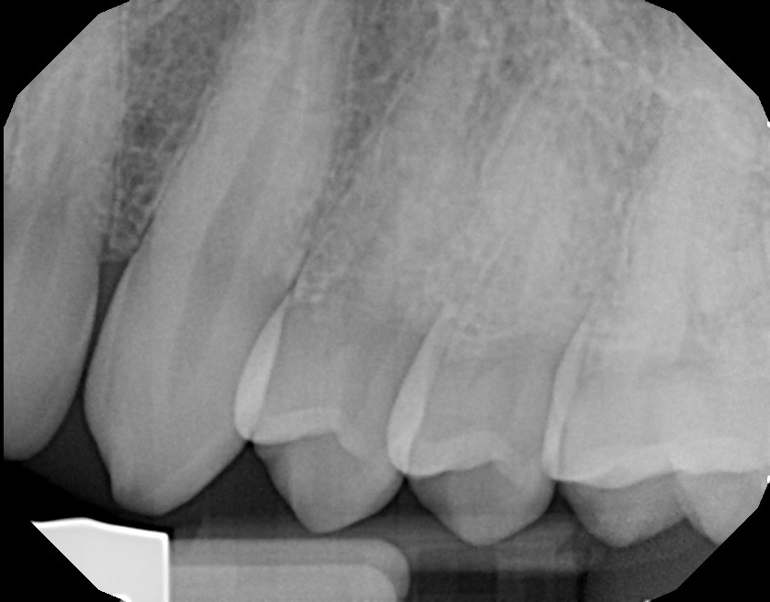
2018 October
From Victoria Olshansky D.D.S. in West Hollywood.
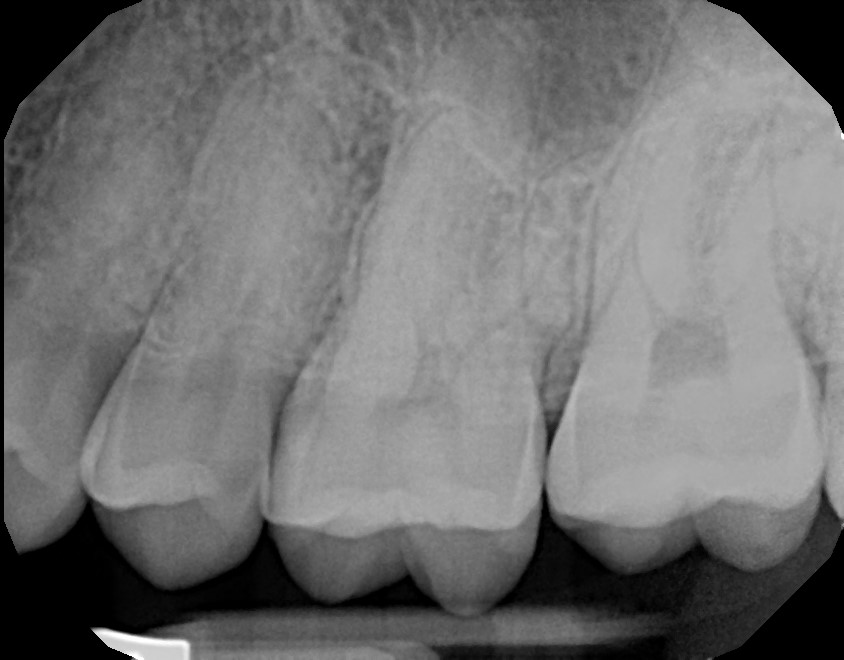
2018 July
From Smile Atelier Beverly Hills Dentist – a super-modern Sunset Strip office.
2017 January
From a hack dentist in West Hollywood who I will refrain from crediting. Note that each image in this collection is separate.
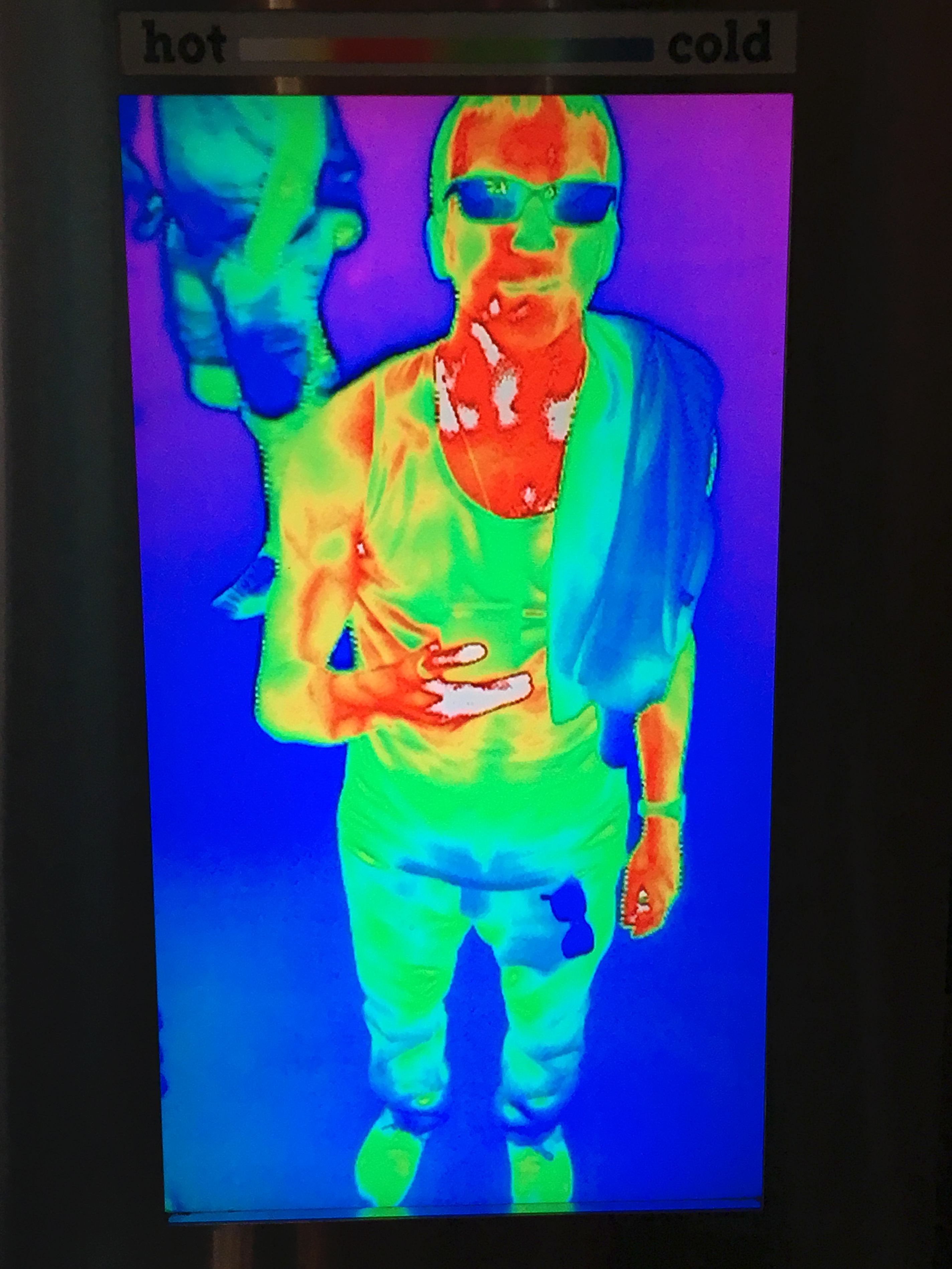
Thermographic
Okay, this one isn’t actually an x-ray, but I think a heat-vision thermal image of my body temperature is close enough to be interesting. This is from a 2017 visit to the LA Natural History Museum.
Thank you for reviewing my portfolio. Now you can say you know me inside and out. Have your bone agent get in touch with my bone agent.